
|
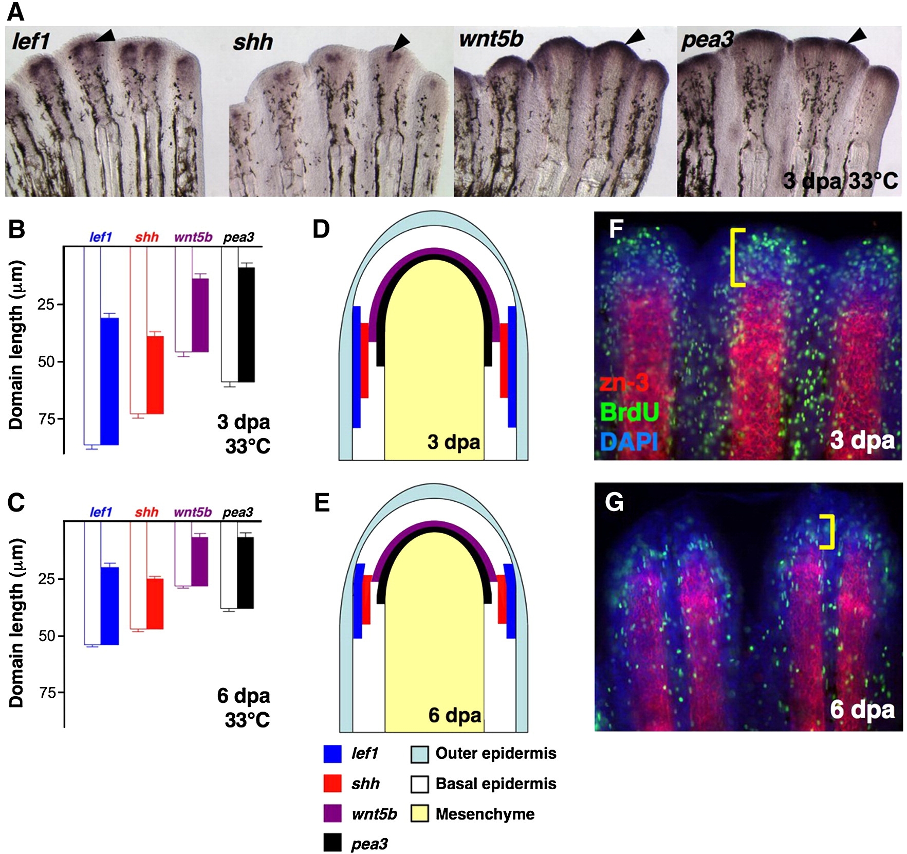
Fig. 1 Expression of epidermal regulators in the regenerating zebrafish fin. (A) Whole-mount ISH of 3 dpa fin regenerates (33 °C) for lef1, shh, wnt5b and pea3 (Black arrowheads, violet). (B,C) Quantification of epidermal expression domains measured from either 3 dpa (B) or 6 dpa (C) whole-mount ISH-processed fins, plotted as mean ± SEM. White bars: length from distal tip of regenerate to proximal end of ISH signal; colored bars: total length of ISH expression domain. (D,E) Cartoon representation of data from (B, C), represented as a longitudinal section of 3 dpa (D) or 6 dpa (E) fin regenerates. (F,G) Whole-mount staining for BrdU (green) and scleroblasts (red) in 3 dpa (F) and 6 dpa (G) fin regenerates. Bracket indicates the proliferative blastema.
Reprinted from Developmental Biology, 331(2), Lee, Y., Hami, D., De Val, S., Kagermeier-Schenk, B., Wills, A.A., Black, B.L., Weidinger, G., and Poss, K.D., Maintenance of blastemal proliferation by functionally diverse epidermis in regenerating zebrafish fins, 270-280, Copyright (2009) with permission from Elsevier. Full text @ Dev. Biol.