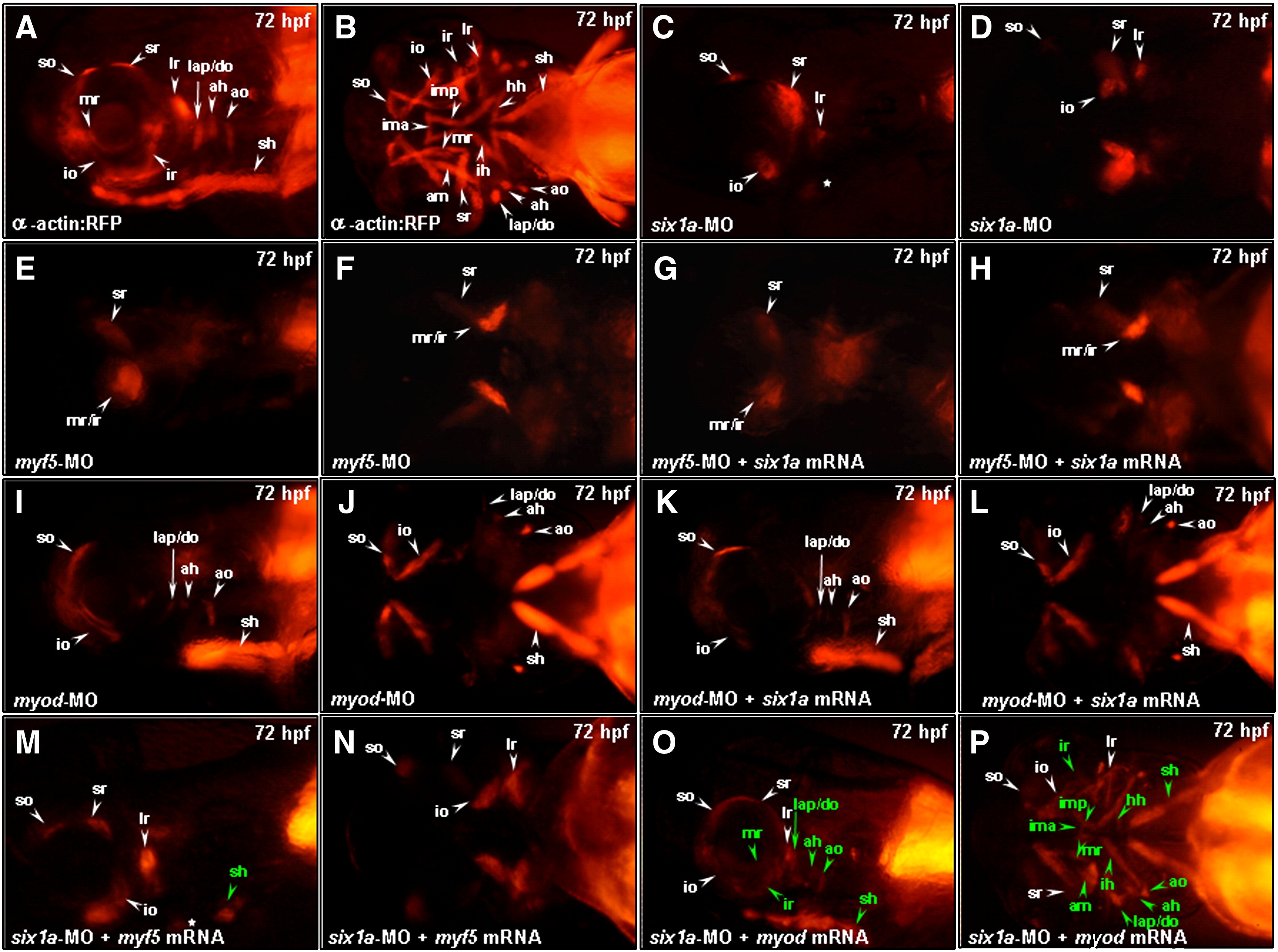
Fig. 4 Injection of myod mRNA enables embryos to rescue the defective muscle derived from cranial mesoderm in the six1a morphants. Embryos derived from Tg (α-actin:RFP) (A, B as non-treated) were injected with 8 ng of six1a-MO (C, D) and either 4 ng of myf5-MO (E, F) or myod-MO (I, J) to serve as control groups. Embryos injected with 4 ng of myf5-MO and 150 pg of six1a mRNA (G, H), 4 ng of myod-MO and 150 pg of six1a mRNA (K, L), 8 ng of six1a-MO and 100 pg of myf5 mRNA (M, N), and 8 ng of six1a-MO and 50 pg of myod mRNA (O, P) were used to examine the appearance of RFP-labeled muscles. Results showed that only sr and mr/ir muscles exhibited in the myf5-MO-injected embryos and in the myf5-MO-six1a-mRNA-injected embryos (E vs. G and F vs. H). In contrast, only so, io, lap/do, ah, ao, and sh muscles exhibited in the myod-MO-injected embryos and in the myod-MO-six1a-mRNA-injected embryos (I vs. K and J vs. L). Similar to six1a morphants, embryos co-injected with six1a-MO and myf5 mRNA exhibited the so, io, sr, lr and remnant dorsal branchial arch muscles. However, injection of myf5 mRNA enabled embryos to rescue only sh primordia muscle among defects induced by six1a-MO (M, N), while injection of myod mRNA enabled embryos to rescue all the defective muscle primordia induced by six1a-MO (O, P). The rescued muscles are marked in green typeface. Lateral view: A, C, E, G, I, K, M and O; Ventral view: B, D, F, H, J, L, N and P. ah, adductor hyoideus; am, adductor mandibulae; ao, adductor operculi; do, dilator operculi; hh, hyohyoideus; ih, interhyoideus; ima, intermandibularis anterior; imp, intermandibularis posterior; io, inferior oblique; ir, inferior rectus; lap, levator arcus palatini; lr, lateral rectus; mr, medial rectus; sh, sternohyoideus; so, superior oblique and sr, superior rectus.
Reprinted from Developmental Biology, 331(2), Lin, C.Y., Chen, W.T., Lee, H.C., Yang, P.H., Yang, H.J., and Tsai, H.J., The transcription factor six1a plays an essential role in the craniofacial myogenesis of zebrafish, 152-166, Copyright (2009) with permission from Elsevier. Full text @ Dev. Biol.

