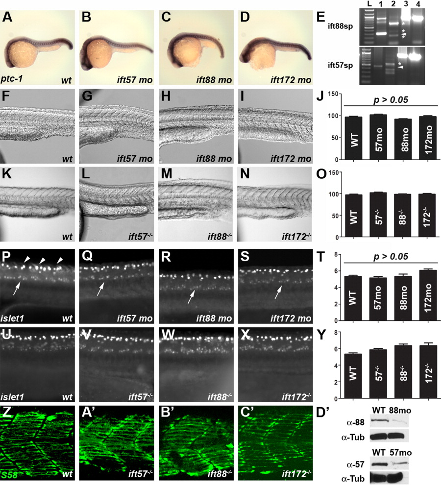
Fig. 9 Loss of IFT function does not affect somite formation or spinal motor neuron development. A-D: Expression of ptc-1 in wild type and IFT morphants is unchanged at 24 hpf. In situ hybridization was done simultaneously and embryos were developed for the same amount of time. At least 20 embryos were examined for each treatment. E: RT-PCR of embryos injected with ift88 (top) or ift57 (bottom) splice-blocking morpholinos. Lane 1 is an uninjected control and shows the wild type transcript, as indicated by the arrowhead in each panel. Lane 2 is RT-PCR from a morpholino injected animal at 72 hpf. Aberrant splice products are denoted by asterisks and no wild type product is present. Injection of ift88-sp morpholinos caused the inclusion of intron 4-5 (340 bp) and resulted in a longer amplification product as the major band. Two minor products likely reflect loss of exon 4 (60 bp) and an aberrant splice product. Injection of ift57-sp morpholinos results in the inclusion of intron 2-3 (88 bp) and results in a longer amplification product and introduces a stop codon. Two smaller amplification products were also observed. All products are predicted to produce frame-shifts and introduce a stop codon. Lanes 3 and 4 show amplification of beta-actin from cDNA used in lanes 1 and 2, respectively. F-O: Brightfield images using Nomarski optics reveal no defects in somites in wild type and IFT morphants or mutants. Somites form the characteristic chevron shape in all IFT morphant and mutant embryos. Quantification of somite angles (J, O) revealed no statistical difference between wild type and IFT morphants or mutants. P-Y: Whole-mount immunocytochemistry with the islet-1 antibody labels motor neurons in the ventral spinal cord (arrows). Clusters of 5-7 positively-stained neurons were typically seen on each side of the embryos. These neurons are found in similar numbers among wild type, IFT mutant, or IFT morphants embryos (T, Y). This antibody also strongly labels Rohon-Beard sensory neurons in the dorsal spinal cord (arrowheads). Z-C′: S58 staining labeled slow-muscle fibers in the chevron-shaped somites of both wild type and genotyped mutant embryos at 24 hpf. Some muscle fibers stained with S58 were below the plane of optical sectioning and not contained in these images. D′: Western blot analysis of 18-hpf embryos injected with IFT morpholinos. Extracts of wild type (wt) and morphant embryos (88mo or 57mo) were immunoblotted for either IFT88 or IFT57 protein. Alpha tubulin (α-Tub) was used as a loading control.
Image
Figure Caption
Figure Data
Acknowledgments
This image is the copyrighted work of the attributed author or publisher, and
ZFIN has permission only to display this image to its users.
Additional permissions should be obtained from the applicable author or publisher of the image.
Full text @ Dev. Dyn.

