Image
Figure Caption
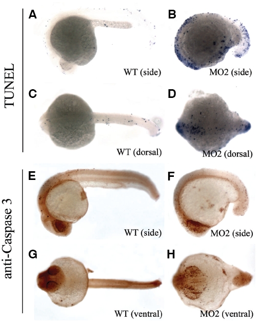
Fig. 5 Apoptosis was increased in mats1 morphant embryos. (A-D) TUNEL staining results at 24 hpf derived from wild type embryos (A,C) and MO2-treated embryos (B,D). (E-H) Cleaved Caspase 3 antibody staining results at the same developmental stage with wild type embryos (E,G) and MO2 treated embryos (F,H). Anterior is towards left in all panels. Both of TUNEL and cleaved Caspase 3 antibody-staining results showed that apoptosis increased in mats1 morphant embryos.
Figure Data
Acknowledgments
This image is the copyrighted work of the attributed author or publisher, and
ZFIN has permission only to display this image to its users.
Additional permissions should be obtained from the applicable author or publisher of the image.
Full text @ Int. J. Dev. Biol.

