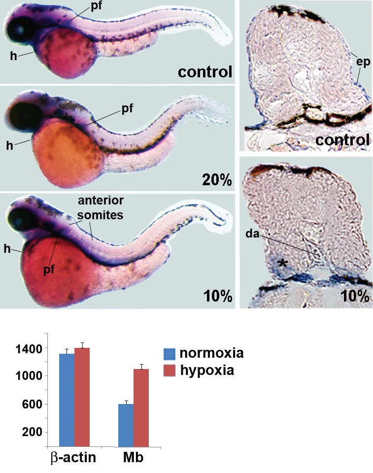
Fig. 4 Influence of hypoxia on myoglobin (Mb) gene expression in zebrafish embryos. Zebrafish embryos were exposed to two different oxygen concentrations (10% and 20% air saturated). Controls were kept at 100% air saturated. Embryos were fixed at 48 hpf for the control (A) and the 20% air saturated water embryos (C) and at 4 dpf for the 10% air saturated water embryos (E) (which were strongly delayed in development). Fixed embryos were subjected to whole mount in situ hybridization. Blue and purple staining indicates myoglobin expression. Similar results were seen in five different specimen and staining in the anterior somites was never observed in control or 20% air saturated water embryos. Staining in the pectoral fins (pf) and in the heart (h) is indicated for all embryos and anterior somites and the dorsal aorta (da) are marked in the 10% air saturated water embryos. In addition, shown are sections of the anterior somite area of control embryos (B) and embryos exposed to 10% air saturated water (D). The latter shows stronger staining in regions of the anterior somites supporting the results of the whole mount pictures. Skeletal muscles are indicated with an asterisk and staining in the dorsal aorta (da) (D). The quantitative RT-PCR results for 48 hpf embryos under normoxic conditions or exposed to hypoxia (10%) are shown in (F). Relative expression of myoglobin is given based on normalization to β-actin. A standard curve for β-actin was included in the experiments. Data represents two independent experiments each done in triplicates. In 48 hpf embryos, 10% hypoxia led to a 1.7 fold induction of Mb normalized to actin (SD± 0.06).
Image
Figure Caption
Figure Data
Acknowledgments
This image is the copyrighted work of the attributed author or publisher, and
ZFIN has permission only to display this image to its users.
Additional permissions should be obtained from the applicable author or publisher of the image.
Full text @ Int. J. Dev. Biol.

