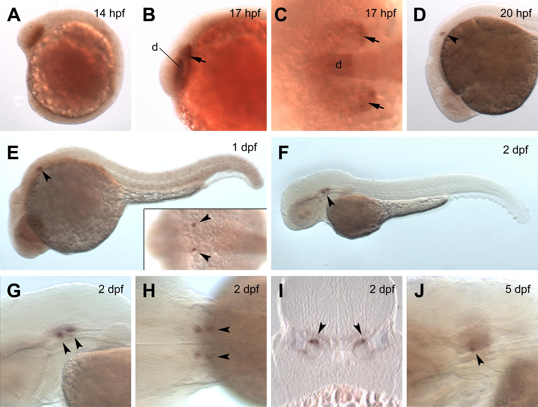
Fig. 4 Analysis of zebrafish foxg1c expression by in situ hybridization. Transcripts are detected in the forebrain and eye primordia of embryos at 14 hpf (A) and 17 hpf (B and C). Expression at 20 hpf is mainly detected in the otic vesicles (D), and similar expression is observed at 1 dpf (E). A dorsal view (inset in E) shows expression restricted to single spots in each otic vesicle (marked by arrowheads). (F) Expression in the otic vesicles is stronger at 2 dpf. Lateral (G) and dorsal (H) views of the larva in (F) at higher magnification show expression in two spots within each of the otic vesicles. (I) Cross-section showing expression restricted to the epithelium in the inner otic vesicle at 2 dpf (orientation with dorsal up). (J) Expression becomes weaker by 5 dpf. Orientations in (A–H, J) are with anterior to the left. Expression signals are indicated with arrowheads (otic vesicles) and arrows (eye primordia). Abbreviations: d, diencephalon; dpf; days post-fertilization; hpf, hours post-fertilization.
Reprinted from Gene expression patterns : GEP, 9(5), Zhao, X.F., Suh, C.S., Prat, C.R., Ellingsen, S., and Fjose, A., Distinct expression of two foxg1 paralogues in zebrafish, 266-272, Copyright (2009) with permission from Elsevier. Full text @ Gene Expr. Patterns

