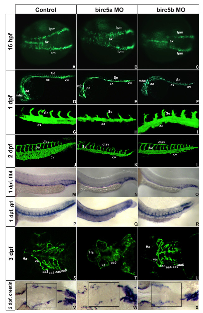
Fig. 3 Birc5 in vasculogenesis and angiogenesis. Tg(Fli:eGFP) (A-L) and Tg(Flk1:GFP) (S-U) embryos. A-C: 16 hpf (14 somites), Birc5a morphants with angioblast migration defects from lateral plate mesoderm (B) which are minor in Birc5b morphants (C). D-F: 1 dpf, Birc5a (E, H) and Birc5b (F, I) morphants have thinner axial vessels and poor caudal vein plexus development. G-I: 1 dpf Birc5a depletion (H) delays intersomitic vessels; not with Birc5b depletion (I). J-L: 2dpf Birc5a morphants with abnormal dorsal longitudinal anastomic and intersomitic vessels. Both morphants have poorly developed caudal vein plexus. M-O: flt4 at 1 dpf is reduced in posterior cardinal vein in both morphants (N, O). P-R: gridlock (grl) at 1 dpf is reduced with Birc5a depletion (Q), but not with Birc5b (R). S-U: 3 dpf Birc5a morphants have hypoplastic aortic arches (T). Birc5b depletion at 3 dpf causes hypoplasia of aortic arches 5–6 (U). V-X: Birc5a depletion decreases neural crest cells that migrate to branchial arches, detected with crestin probe. Birc5b depletion (L) reduces neural crest cells. ax: axial vessels, mhc: midbrain-hindbrain channel, Se: intersomitic vessels, aa: aortic arch, cv: caudal vein plexus, pcv: posterior cardinal vein, da: dorsal aorta, dlav, dorsal longitudinal anastomic vessel, Ha: hypobranchial artery, va: ventral aorta, lpm: lateral plate mesoderm.
Image
Figure Caption
Figure Data
Acknowledgments
This image is the copyrighted work of the attributed author or publisher, and
ZFIN has permission only to display this image to its users.
Additional permissions should be obtained from the applicable author or publisher of the image.
Full text @ BMC Dev. Biol.

