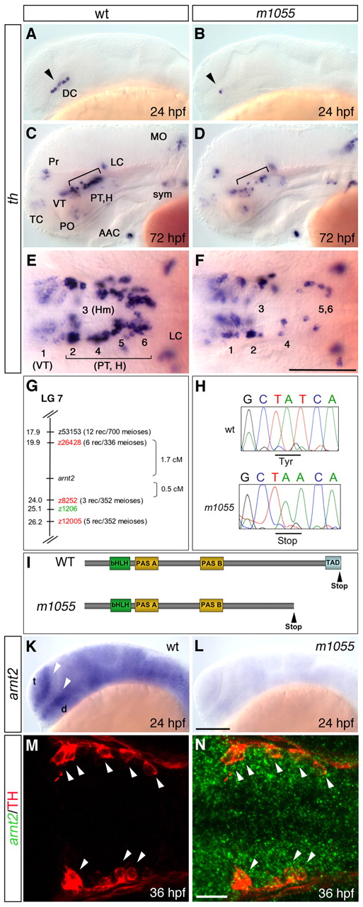
Fig. 1 A mutation in arnt2 affects DA neuron groups in the ventral diencephalon of zebrafish. (A-F) Reduction of th-expressing DA neuron groups in the ventral diencephalon of m1055 mutants (B,D,F) compared with wild-type siblings (A,C,E) at 24 hpf (A,B) and 72 hpf (C-F). (G) Genetic mapping of the m1055 allele. SSLP markers (MGH panel black and T51 panel red) and genetic distances are shown. (H) Sequencing of arnt2 in m1055 mutants reveals a T to A exchange at position 1869. (I) Protein structure of the PAS family member arnt2. The premature stop codon in the m1055 allele disrupts the essential transactivation domain (TAD). (K,L) arnt2 is broadly expressed in wild type (K) at 24 hpf, but almost absent in m1055 mutants (L). (M,N) Fluorescent whole-mount in situ hybridization for arnt2 (green) combined with immunohistochemistry for TH (red) at 36 hpf. Arrowheads indicate co-expression. (A-D,K-L) Lateral views, (E,F,M,N) dorsal views, anterior towards the left. Scale bars: in F, 100 μm for E,F; in L, 100 μm for A-D,K,L; in N, 25 μm for M,N. AAC, arch associated cluster; d, diencephalon; DC, diencephalic cluster; H, hypothalamus; Hm, medial hypothalamus; LC, locus coeruleus; MO, medulla oblongata; PO, preoptic region; Pr, pretectum; PT, posterior tuberculum; sym, sympathetic CA neurons; t, telencephalon; TC, telencephalic cluster; VT, ventral thalamic cluster. Numbers in E,F indicate DA clusters in VT (1) and PT/H (2-6) according to Rink and Wullimann (Rink and Wullimann, 2002).
Image
Figure Caption
Figure Data
Acknowledgments
This image is the copyrighted work of the attributed author or publisher, and
ZFIN has permission only to display this image to its users.
Additional permissions should be obtained from the applicable author or publisher of the image.
Full text @ Development

