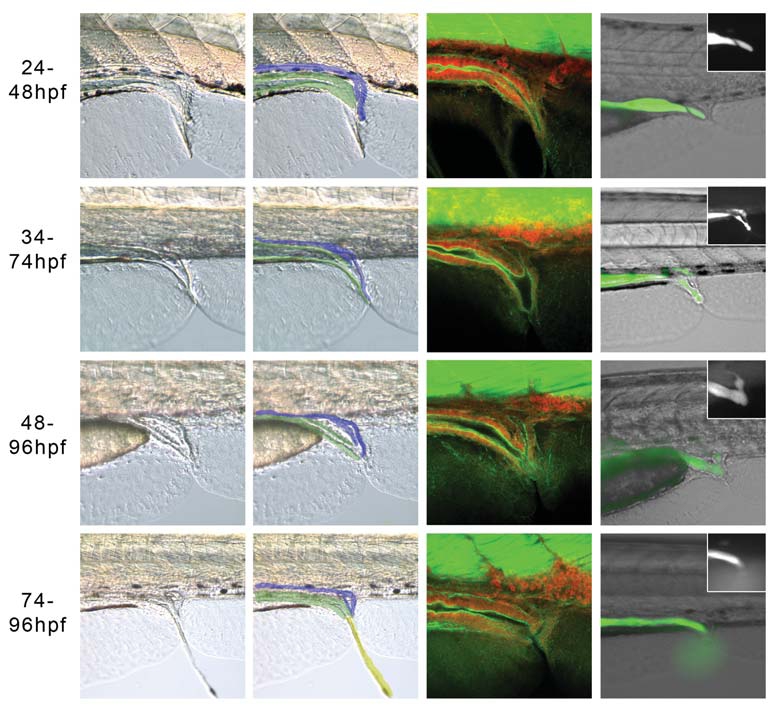
Fig. 7 Hh signalling is required between 48-74 hpf for the development of a perforate anus. Embryos treated with 25 μM cyclopmine and imaged live at 120 hpf with bright field illumination, (A,B,E,F, I,J,M,N) or by confocal microscopy following staining with α-actin-phalloidin (green) and propidium iodide (red) (C,G,K,O). The overlay in (B,F,J,N) shows the approximate identity of the relevant tissues, with the posterior gut in green and pronephric ducts in blue. (D,H,L,P) Merged brightfield/fluorecent images of live embryos injected with fluorescein salt at 72-96 hpf, with the fluorescent channel only shown in the inset. (A,B) After treatment from 24-48 hpf, the posterior gut is thinner than in WT embryos, and despite the appearance of a fistula (C), the majority of embryos have a perforate gut, while 33% (4/12) were imperforate and the dye stayed in the blind ended posterior gut (D). (E-H) 34-74 hpf treatment. The posterior gut formed a fistula with the pronephric duct in most embryos, which allowed excretion through a single opening in the proctodeum in around half the embryos (10/18). In the embryos that were not perforate, fluorescein entered the pronephros, but could not be excreted and so moved anteriorly through the pronephric tubes, due to peristaltic pressure (H). (I-L) 48-74hpf treatment. Embryos from this treatment window had a range of phenotypes, 67% (16/24) were perforate, but around half had a fistula to the pronephros (14/24), which itself was not always perforate leading to fluorescein in the pronephric ducts (L). Other embryos had atresia or stenosis. (M-P) 74-96 hpf treatment. The majority of embryos are perforate (90%, 18/20) and the posterior gut musculature affords some control over the excretion of faecal matter. (P) Faecal matter being expelled in a live embryo.
Image
Figure Caption
Acknowledgments
This image is the copyrighted work of the attributed author or publisher, and
ZFIN has permission only to display this image to its users.
Additional permissions should be obtained from the applicable author or publisher of the image.
Full text @ Int. J. Dev. Biol.

