Image
Figure Caption
Fig. S8
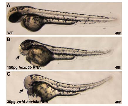
Embryos Injected with hoxb5b RNA or vp16-hoxb5b RNA Display Similar Phenotypes
(A–C) Lateral views, anterior to the left, of live embryos at 48 hpf.
(A) WT embryo.
(B) Embryo injected with 150 pg of hoxb5b RNA.
(C) Embryo injected with 30 pg of vp16-hoxb5b RNA. Both hoxb5b RNA and vp16- hoxb5b RNA posteriorize the embryo, resulting in the loss of anterior structures, like the eyes (arrows in B and C). However, vp16-hoxb5b RNA is more potent and can achieve the same phenotypes as hoxb5b RNA at one-fifth the dose.
Acknowledgments
This image is the copyrighted work of the attributed author or publisher, and
ZFIN has permission only to display this image to its users.
Additional permissions should be obtained from the applicable author or publisher of the image.
Reprinted from Developmental Cell, 15(6), Waxman, J.S., Keegan, B.R., Roberts, R.W., Poss, K.D., and Yelon, D., Hoxb5b acts downstream of retinoic Acid signaling in the forelimb field to restrict heart field potential in zebrafish, 923-934, Copyright (2008) with permission from Elsevier. Full text @ Dev. Cell

