Image
Figure Caption
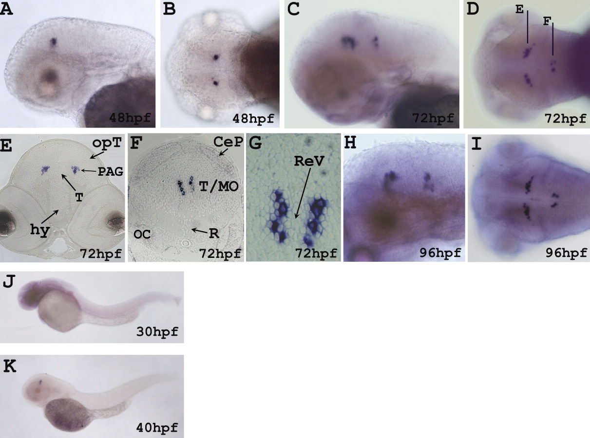
Fig. 1 Whole mount in situ hybridization of rln3 on embryos at indicated stages. Lateral and dorsal view of the zebrafish brain at 48 hpf (A, B), 72 hpf (C, D), and 96 hpf (H, I). E,F: Transverse sections indicated by the black lines in D. G: A magnification of a transverse section centred at the level of NI. Lateral view showing the expression of rln3 gene in the zebrafish brain at 30 and 40 hpf (J, K). CeP, cerebellar plate; hy, hypothalamus; MO, medulla oblongata; OC, otic capsule; opT, optic tectum; PAG, periaqueductal gray; R, raphe; ReV, rombencephalic ventricle; T, tegmentum.
Figure Data
Acknowledgments
This image is the copyrighted work of the attributed author or publisher, and
ZFIN has permission only to display this image to its users.
Additional permissions should be obtained from the applicable author or publisher of the image.
Full text @ Dev. Dyn.

