
|
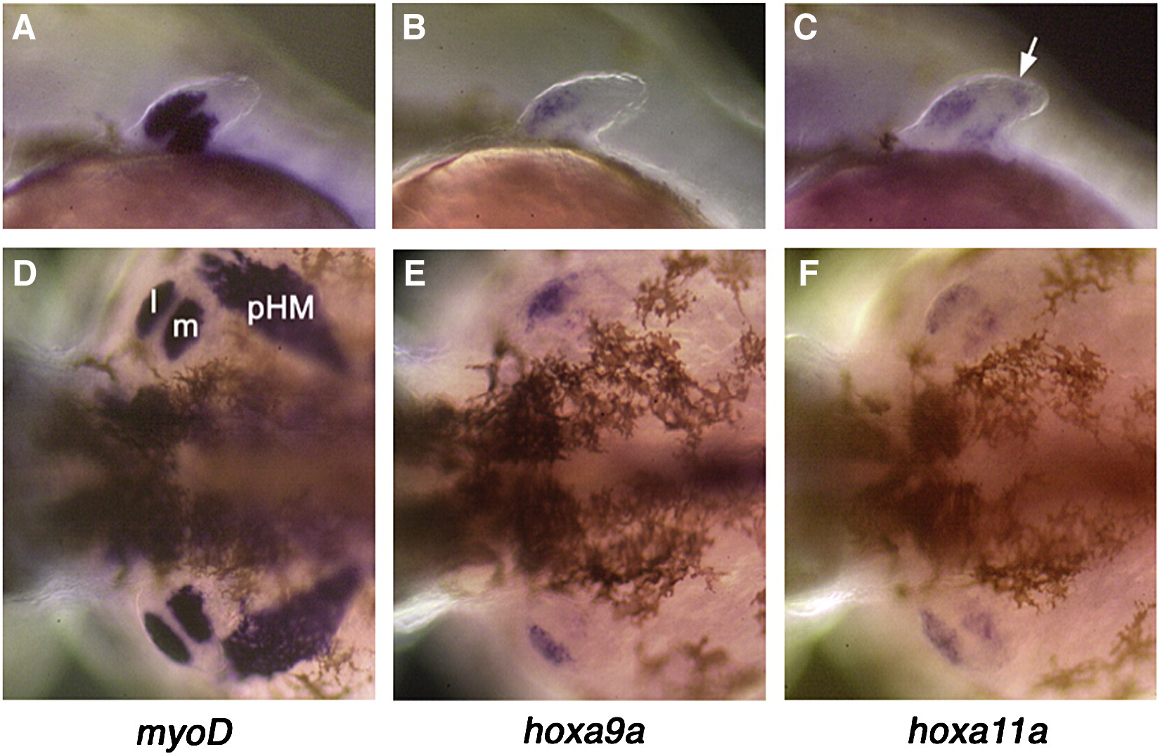
Fig. S1 Late expression of hoxa9a/a11a in pectoral fin muscle cells in zebrafish. By 48 hpf, pectoral fin muscle cells can be recognized by the expression of several marker genes such as myoD (A, D), which is not expressed during the migration of fin muscle cells (24–30 hpf: not shown). At this stage, it is clear that the expression of hoxa9a (B, E) and hoxa11a (C, F) is mostly confined to the pectoral fin muscle cells. Note that, for hoxa9a, expression is much weaker in the medial cluster of muscle cells compared to the lateral cluster of muscle cells. For hoxa11a, expression is also seen in the distal mesenchyme cells (arrow in B) in addition to the pectoral fin muscle cells. A–C: lateral view of left pectoral fins. D–F: dorsal-posterior view of pectoral fins and surrounding regions. l: lateral cluster of fin muscle cells. m: medial cluster of fin muscle cells. pHM: posterior hypaxial muscle.
Reprinted from Developmental Biology, 322(1), Ahn, D., and Ho, R.K., Tri-phasic expression of posterior Hox genes during development of pectoral fins in zebrafish: Implications for the evolution of vertebrate paired appendages, 220-233, Copyright (2008) with permission from Elsevier. Full text @ Dev. Biol.