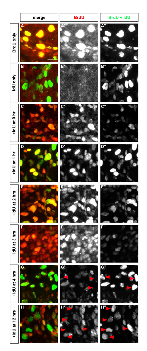
Fig. S1 BrdU remains available for 4 hours after injection. To measure the perdurance of BrdU in zebrafish embryos, BrdU was chased with IdU. Embryos were injected with BrdU and/or IdU and were fixed 2 hours after the last injection. Embryos were stained with an antibody that recognized BrdU only (AbD Serotec) and an antibody that recognized both BrdU and IdU (Becton Dickinson). Primary antibodies were detected using an anti-rabbit antibody coupled with rhodamine and an anti-mouse antibody coupled with FITC (Jackson ImmunoLab), respectively. Therefore, cells labeled with BrdU only or with BrdU and IdU have a red and green signal (yellow in merged image), whereas cells that are labeled with IdU only have a green signal. (A-C) Staining of embryos injected with BrdU only (A), IdU only (B), or both (C), revealed that the anti-BrdU antibody specifically recognized BrdU, whereas the anti-BrdU/IdU antibody recognized both nucleotides. (D-H) Embryos were injected first with BrdU at 24 hpf and injected again with IdU 1 hour (D), 2 hours (E), 3 hours (F), 4 hours (G) and 12 hours (H) following the BrdU injection. When IdU was injected 4 hours after the BrdU injection, several green stained cells were visible throughout the embryo, indicating that BrdU is no longer available to be incorporated 4 hours after injection. Side view, anterior towards the left.
Image
Figure Caption
Figure Data
Acknowledgments
This image is the copyrighted work of the attributed author or publisher, and
ZFIN has permission only to display this image to its users.
Additional permissions should be obtained from the applicable author or publisher of the image.
Full text @ Development

