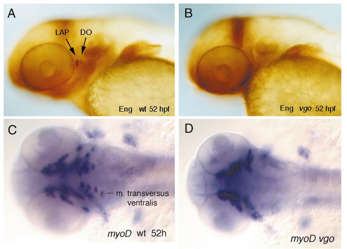
Fig. 13 Whole-mount in situ hybridization experiments with genes that are expressed in the mesoderm. (A and B) Lateral views of two jaw muscles, the levator arcus palatini (LAP) and the dilator operculi (DO), that express Engrailed during development. (B) In 52-hpf vgo embryos these two muscles are absent, as revealed by antibody labeling with 4D9, which recognizes Eng. All other voluntary pharyngeal muscles express myoD. (C and D) Ventral views of myoD expression in 52-hpf wild-type (C) and vgo (D) embryos. In vgo embryos the muscles which are associated with the pharyngeal arches 4–7 do not express myoD or are absent.
Reprinted from Developmental Biology, 225(2), Piotrowski, T. and Nüsslein-Volhard, C., The endoderm plays an important role in patterning the segmented pharyngeal region in zebrafish (Danio rerio), 339-356, Copyright (2000) with permission from Elsevier. Full text @ Dev. Biol.

