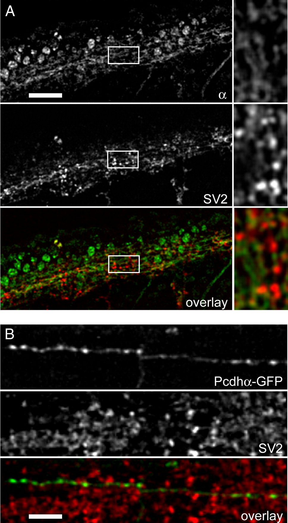
Fig. 5 Pcdh1α does not colocalize with presynaptic markers. (A) Maximum intensity projections of 3 adjacent optical sections (spacing is 1 μm) were generated in order to reconstruct an extended region of labeled axon tracts in the spinal cord of a 24 hpf embryo. The top panel shows labeling with antibody against Pcdhα; the middle panel shows staining with the presynaptic marker, SV2; the bottom panel shows the overlay. The panels on the right are magnified views of the boxed regions. Although both SV2 and Pcdh1α antibodies label many of the same axons, the staining patterns are distinct with Pcdhα failing to exhibit punctate staining that overlaps with SV2. Scale bar = 25 μm. (B) Shown are maximum intensity projections of 2 adjacent optical sections (spacing is 0.5 μm). The top panel shows a segment of axon labeled with Pcdhα-GFP, which was then processed for whole-mount immunocytochemistry. In general, the labeling of Pcdhα-GFP in fixed tissue appears more discontinuous than in living embryos. The middle panel shows the same field, which has been labeled with SV2 antibodies. Although the discontinuity of Pcdhα-GFP labeling has an almost punctate appearance, it does not significantly overlap, or co-vary, with the punctate SV2 labeling. Scale bar = 8 μm.
Reprinted from Developmental Biology, 321(1), Emond, M.R., and Jontes, J.D., Inhibition of protocadherin-alpha function results in neuronal death in the developing zebrafish, 175-187, Copyright (2008) with permission from Elsevier. Full text @ Dev. Biol.

