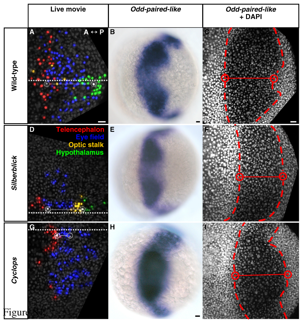
Fig. S1 Initiation of keel formation relative to eye-field location in wild-type, silberblick and cyclops embryos. (A-C) Wild type. (D-F) slb. (G-I) cyc morphant. (A-I) Dorsal views, anterior to left. (A,D,G) Forebrain regions, defined by cell tracking, at 80% epiboly (8.4 hpf). White cells and open circles - medial anterior eye (left) and initiation of keel formation (right). Adjoining solid line - intervening distance along the midline (square dotted line). Keel formation begins with hypothalamus in wild-type (A) and slb (D), and with anterior eye field in cyc morphant (G). For corresponding angular distances subtended at the embryonic centroid see Table S1. (B,E,H) Odd-paired-like (opl) gene expression, as determined using standard procedures, defines the eye field at 80% epiboly (8.4 hpf). Hypothalamus occupies the posterior-medial notch in wild type (B) and slb (E), consistent with the tracked data in A and D. However, eye tissue presides in the posterior-medial notch in cyc morphant (H). (C,F,I, Table S1) DAPI nuclear counter-staining, defining the medial boundaries (red cells and open circles) of the opl-expressing eye field (dashed line) at 80% epiboly (8.4 hpf), confirms this. hpf, hours post fertilisation. Scale bars: 25 μm.
Image
Figure Caption
Figure Data
Acknowledgments
This image is the copyrighted work of the attributed author or publisher, and
ZFIN has permission only to display this image to its users.
Additional permissions should be obtained from the applicable author or publisher of the image.
Full text @ Development

