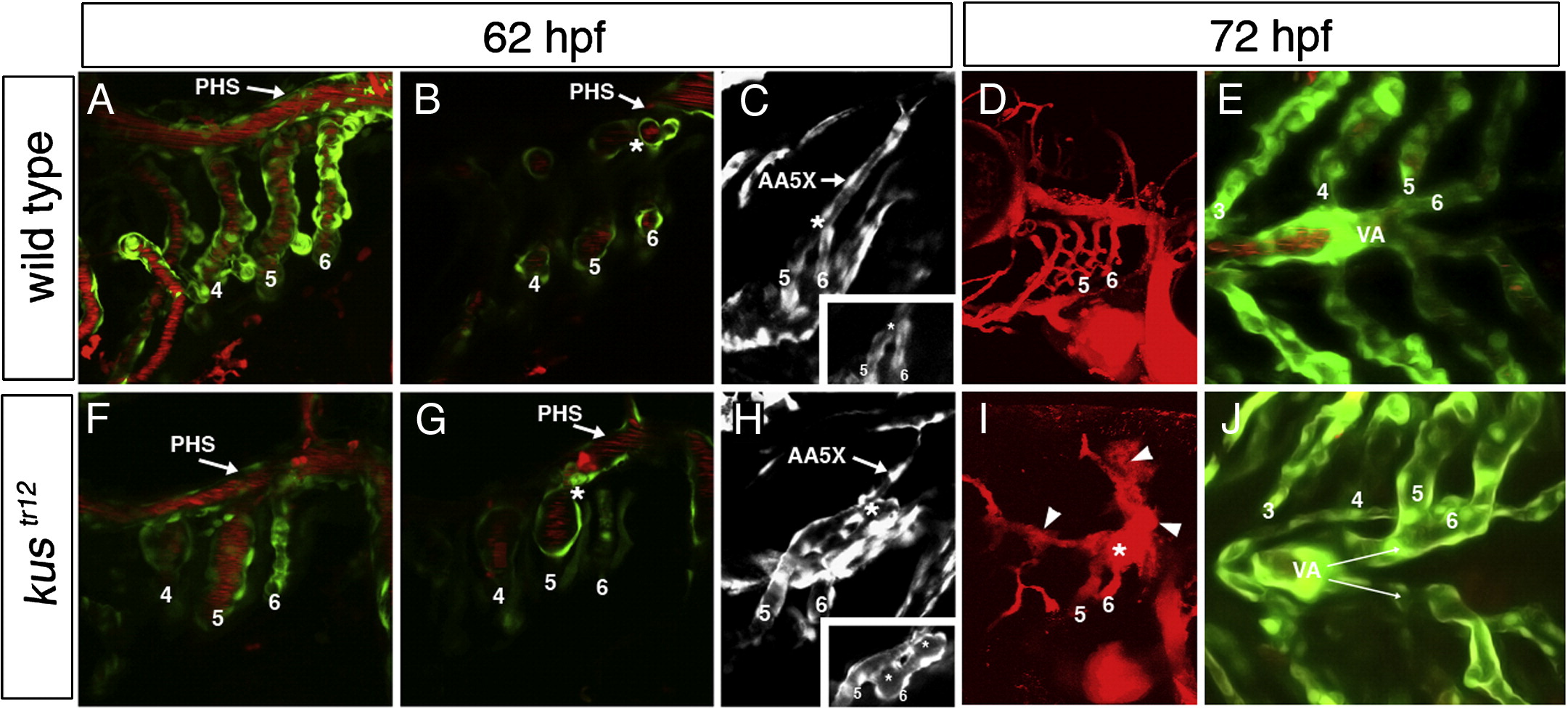
Fig. 3 Arteriovenous malformation and ventral aorta defect in kustr12 mutants. In wild type embryos, aortic arches 5 and 6 fuse just behind the primary head sinus (PHS) and connect to the lateral dorsal aorta via an extension of aortic arch 5 (AA5X); clear demarcation can be seen between the aortic arch 5/6 connection (asterisk) and the PHS (A–C). In contrast, in kustr12 mutants, AA5X is poorly formed, and the enlarged aortic arch 5/6 connection fuses to the adjacent PHS, creating an AVM (asterisk) that shunts blood directly back to the heart (F–H). This AVM carries the majority of blood flow and is typically associated with localized hemorrhage [arrowheads; compare D (WT) to I (kustr12)]. In wild type embryos, the paired ventral aortae (VA) fuse by 72 hpf (E). In contrast, in kustr12, the ventral aortae fail to fuse properly and remain paired at 72 hpf, similar to wild types at 48 hpf (J; compare also to Fig. 1M). 2D confocal projections of TG(flk1:GFPla116;gata1:dsRed) embryos (A, E, F, J); TG(flk1:GFPla116) embryos (C, H); or microangiograms (D, I). Panels B, G and insets in panels C, H are single planes from confocal Z-stacks represented by A, F, C, and H, respectively. A–C; F–H: 62 hpf. D, E, I, J: 72 hpf. A, B, D, F, G, I: lateral views, anterior to the left. C, H: dorsolateral views, anterior to the left. E, J: ventral views, anterior to the left.
Reprinted from Developmental Biology, 318(2), Anderson, M.J., Pham, V.N., Vogel, A.M., Weinstein, B.M., and Roman, B.L., Loss of unc45a precipitates arteriovenous shunting in the aortic arches, 258-267, Copyright (2008) with permission from Elsevier. Full text @ Dev. Biol.

