
|
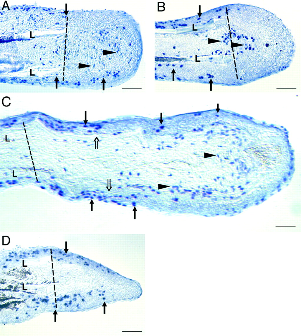
Fig. 5 BrdUrd-incorporation in fins treated with cyclopamine or solanidine. After 1 day of treatment (initiated at 2 dpa) with either solanidine (A) or cyclopamine (B), proliferating cells were identified in the epidermis (solid arrows) and mesenchyme (arrowheads) in the region of new bone matrix secretion. By day 3, BrdUrd-labeled cells were still found in the epidermal layers of both control and cyclopamine-treated fins (C, D, solid arrows) and in the mesenchyme of solanidine-treated fins (C, arrowheads), but not in the mesenchyme of cyclopamine-treated fins (D). Dotted lines mark the level of amputation. (Bar = 50 μm.)