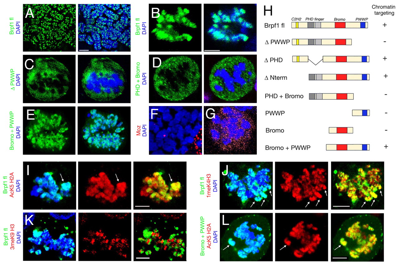
Fig. 6 The PWWP domain is required for association of Brpf1 with metaphase chromosomes. (A-G) Immunofluorescent staining of mitotic HEK 293 cells transfected with the indicated GFP-Brpf1 constructs (A-E; green) and FLAG-Moz (F,G; red). (A,F) Spreads of metaphase chromosomes. Right panels of A-E and F,G show merged images with DAPI staining of DNA (blue). Full-length Brpf1 displays punctate distribution along metaphase chromosomes (A), whereas in intact nuclei, localization is concentrated in fewer, but still distinct domains of the DNA (B). Truncated Brpf1 lacking the PWWP domain (C) and a Brpf1 fragment containing the PHD domain and the bromodomain (D) are excluded from mitotic chromosomes, whereas a Brpf1 fragment containing the bromodomain and the PWWP domain co-localizes with DNA (E) in a similar manner to full-length Brpf1 (B). (F,G) In contrast to Brpf1 (A,B), no chromatin association is apparent for Moz in metaphase chromosome spreads (F) and in intact mitotic nuclei (G). (H) Schematic structures and chromosome-targeting properties of the full-length and truncated versions of Brpf1. (I-L) Immunofluorescent staining of mitotic HEK 293 cells, revealing that full-length Brpf1 (I-K; green) and the fragment containing the bromodomain and PWWP domain (L; green) co-localize with the active chromatin markers H2AK5Ac (I,L; red) and H3K4me1 (J; red), but not with the inactive chromatin marker H3K9me3 (K; red). Left panels are counterstained with DAPI (blue); merged images are shown in right-hand panels; regions with strong co-localization (yellow) are indicated by arrows. Scale bars: 2.5 μm in A; 5 μm in B,I-L.
Image
Figure Caption
Acknowledgments
This image is the copyrighted work of the attributed author or publisher, and
ZFIN has permission only to display this image to its users.
Additional permissions should be obtained from the applicable author or publisher of the image.
Full text @ Development

