Fig. 1
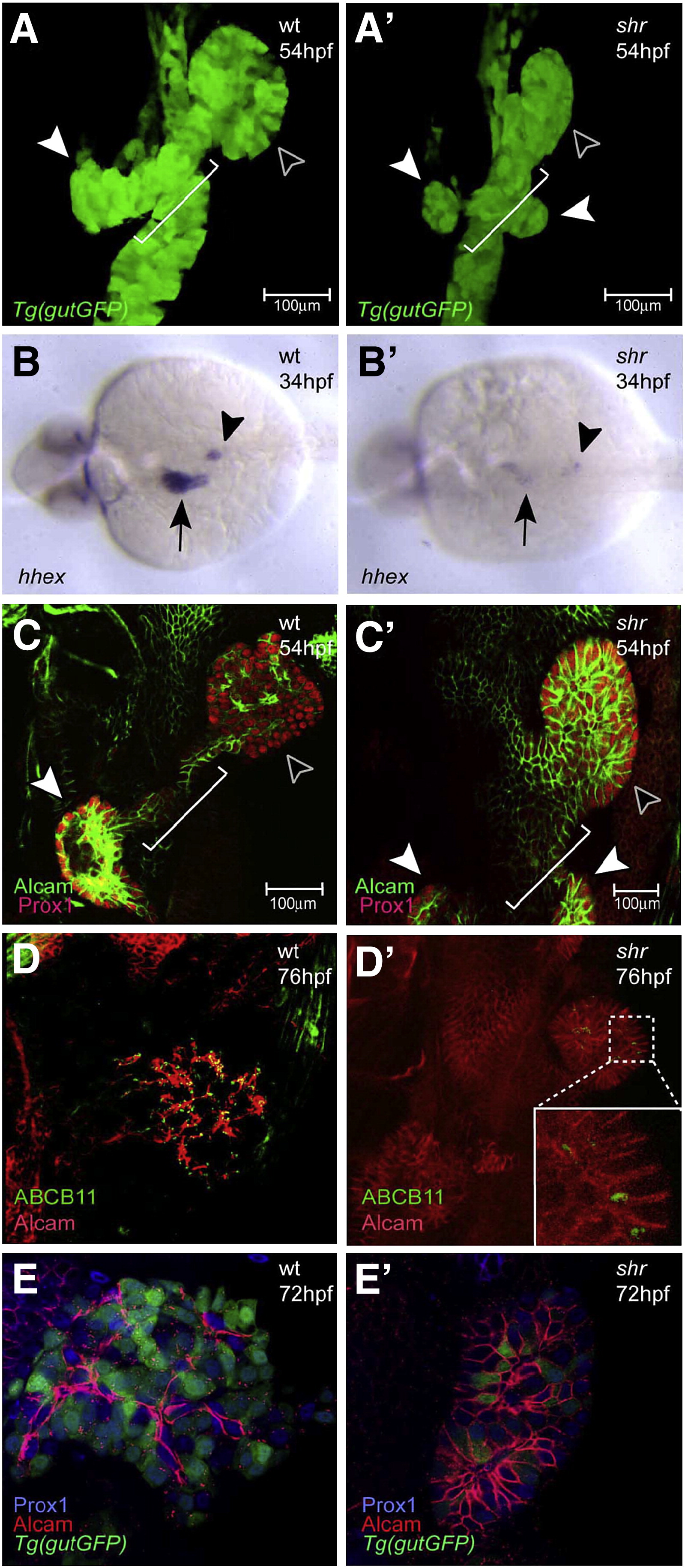
Fig. 1 shr is required for liver and ventral pancreas development. Confocal projections of wild-type and shrs435 mutant embryos in the Tg(gutGFP)s854 line at 54 hpf (A, A′), hhex expression in wild-type and shrs435 mutant embryos at 34 hpf (B, B'), and confocal projections of Alcam (green) and Prox1 (red) expression at 54 hpf (C, C'), of Alcam (red) and ABCB11 (green) expression at 76 hpf (D, D'), and of Alcam (red) and Prox1 (blue) expression in the Tg(gutGFP)s854 line at 72 hpf (E, E′). (A, A′) The size of the liver (black arrowhead) and pancreas (white arrowhead) is often reduced in shrs435 mutants compared to wild-type. By 54 hpf, the formation of the hepatopancreatic duct (white bracket) is initiated in wild-type embryos, whereas shrs435 mutants exhibit an ambiguous duct morphology (white bracket) and two ventral pancreatic buds instead of one (white arrowheads). (B, B′) hhex is clearly expressed in the wild-type liver (black arrow) and pancreatic islet (black arrowhead) while its expression can be detected in only a few cells in the liver and pancreas regions of shrs435 mutants. (C, C′) Prox1 is expressed in the liver (black arrowhead) and pancreas (white arrowhead) in wild-type and shrs435 mutant embryos. At 54 hpf, Alcam has started to show restricted localization in wild-type liver, while it remains distributed along the entire surface of liver cells in shrs435 mutants. To better visualize hepatic Alcam expression in the mutants, a 1.2x magnified image is shown in panel C′. (D, D′) ABCB11 expression is almost absent in shrs435 mutants. To better visualize hepatic ABCB11 expression in the mutants, a magnified image is shown in an inset. (E, E') By 72 hpf, Alcam localizes on or near the apical side of hepatocytes in wild-type, while it remains distributed along the entire surface of liver cells in shrs435 mutants. All images except B, B′ are ventral views, anterior to the top; B, B′ are dorsal views, anterior to the left.
Reprinted from Developmental Biology, 317(2), Shin, C.H., Chung, W.S., Hong, S.K., Ober, E.A., Verkade, H., Field, H.A., Huisken, J., and Stainier, D.Y., Multiple roles for Med12 in vertebrate endoderm development, 467-479, Copyright (2008) with permission from Elsevier. Full text @ Dev. Biol.

