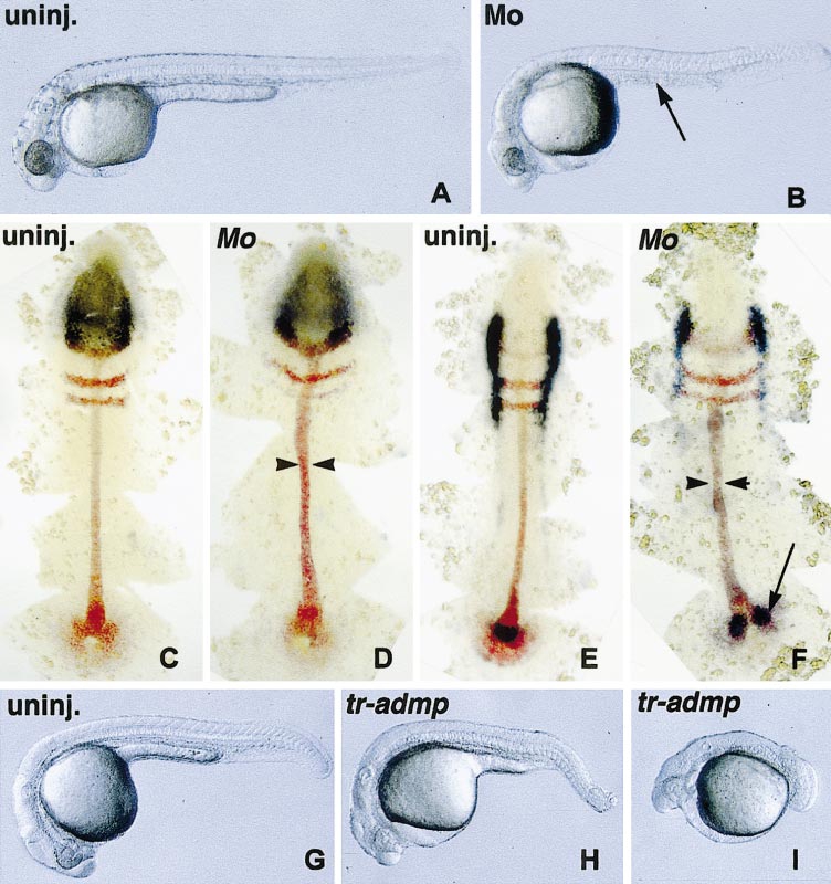
Fig. 6 admp-morpholino and tr-admp RNA promote the expansion of dorso-axial structures. wt embryos were injected at the one-cell stage with admp-morpholino (Mo) (250 μM) or tr-admp RNA (600 pg) and compared to uninjected siblings (uninj.). Embryos injected with a control morpholino were unaffected (not shown) and similar to uninjected siblings. (A, B, G–I) Live embryos at 1 day of development. (A) Uninjected embryo. (B) Mo-injected embryos lack the yolk-tube extension (arrow). (C, D) Flat mounts of embryos stained at the three-somite stage with her5, krox20, ntl probes in red and otx2 probe in dark blue. (E, F) Flat mounts of embryos stained at the three-somite stage with her5, krox20, ntl probes in red and fkd6 probe in dark blue. (D, F) Mo-injected embryos have an enlarged notochord (arrowheads). (F) End of the notochord is split in Mo-injected embryos (arrow). (G) Uninjected embryo. (H) tr-admp-injected embryo, A2 phenotype. (I) tr-admp-injected embryo, A3 phenotype. Note the loss of ventral tail fin and vein in tr-admp-injected embryos that is not observed upon Mo injection.
Reprinted from Developmental Biology, 241(1), Willot, V., Mathieu, J., Lu, Y., Schmid, B., Sidi, S., Yan, Y.-L., Postlethwait, J.H., Mullins, M., Rosa, F., and Peyriéras, N., Cooperative action of ADMP- and BMP-mediated pathways in regulating cell fates in the zebrafish gastrula, 59-78, Copyright (2002) with permission from Elsevier. Full text @ Dev. Biol.

