
|
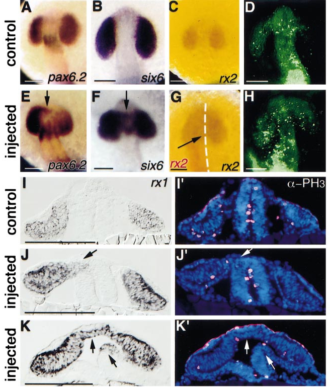
Fig. 8 Ectopic expression of eye-determination genes, cell death, and proliferation in rx2-injected embryos. (A-C) Normal expression patterns. (E–G) Ectopic expression of eye-determination genes (arrows). Note in (C) and (G), purple/brown staining shows the endogenous rx2 expression (detected by a probe transcribed from 3′ coding and 3′ UTR sequences that were not present in the injection construct, rx2DC9). In (G), red staining shows the exogenous rx2 (detected by a probe transcribed from rx2ΔC′). (D, H) Dorsal view of living embryos stained with acridine orange. (H) Note more cell death on the left side of the injected embryo. (I-K′) Transverse sections of embryos showing expression of rx1 (I, J, K) and Cy-3 (red fluorescence) staining of mitotic cells and DAPI nuclear staining (I′, J′, K′) of the same sections, respectively. The arrows in (J-K′) indicate ectopic expression of rx1 in forebrain territory in injected embryos. Embryos were injected with 300 pg β-galactosidase (D), 1 ng rx2ΔC′ (E, G), 300 pg rx2ΔRx (H, J-K′), or 600 pg myc-rx2 (F) RNA, and examined at 16 hpf (A-B, E-F), 15 hpf (C, G), or 19 hpf (D, H, I-K′). Rostral is up in (A-H). α-PH3, anti-phosphohistone H3 antibody. Scale bars: 100 μm.
Reprinted from Developmental Biology, 231, Chuang, J.-C. and Raymond, P.A., Zebrafish genes rx1 and rx2 help define the region of forebrain that gives rise to retina, 13-30, Copyright (2001) with permission from Elsevier. Full text @ Dev. Biol.