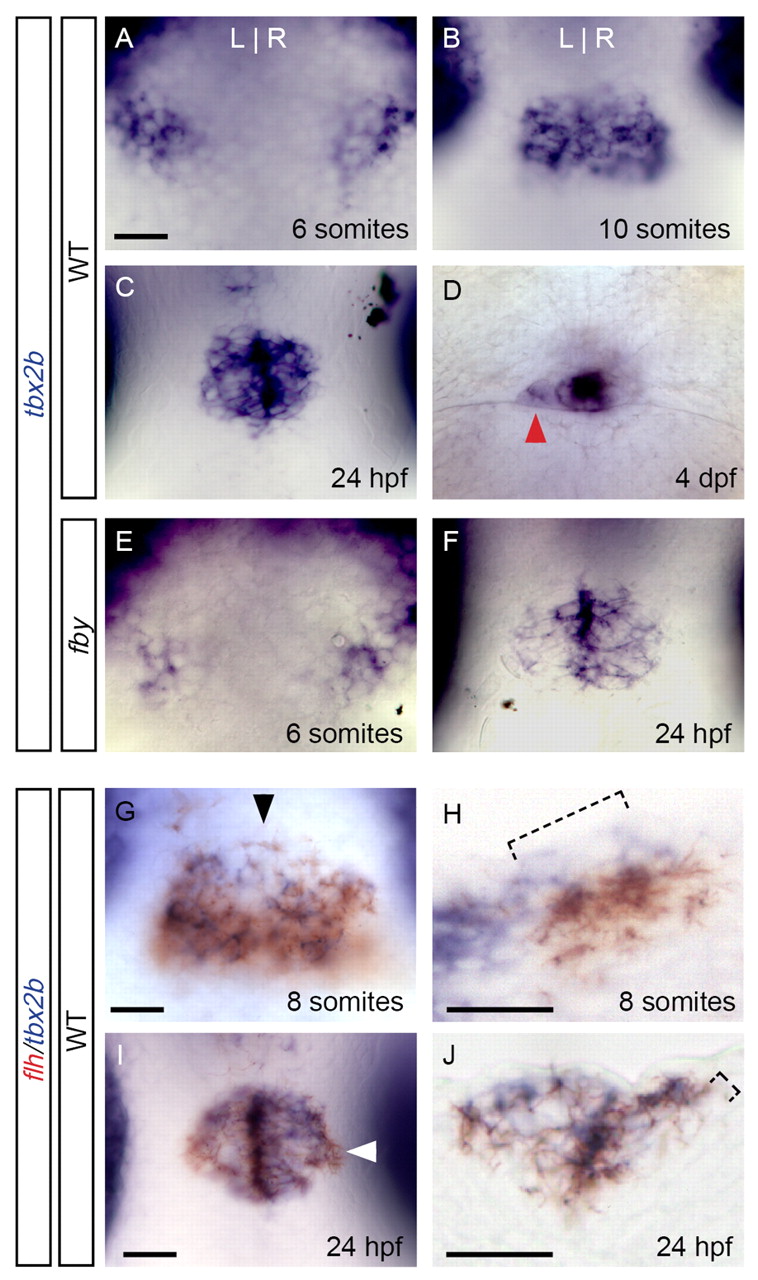
Fig. 7 tbx2b expression in the pineal complex. (A-G,I) Dorsal views of zebrafish embryos or larvae at the indicated stages of development. (H) A sagittal section at the level of the arrowhead in G. (J) A transverse section at the level of the arrowhead in I. (A) Expression of tbx2b in the future epithalamus began at six somites, in two groups of cells at the lateral borders of the neural plate. (B,C) Following neural tube closure, a single tbx2b-expressing domain was detected at ten somites (B) and 24 hpf (C). (D) At 4 dpf, expression was maintained in the parapineal (arrowhead) and in the pineal stalk. (E,F) Expression of tbx2b was unchanged in fby mutants at ten somites (E) and 24 hpf (F). (G,H) Many tbx2b-expressing cells (blue) also expressed flh (red) in the future epithalamus at eight somites (G), although some tbx2b+ flh- cells were detected medially and dorsally (H, bracket). (I,J) By 24 hpf, tbx2b and flh were co-expressed in much of the pineal complex anlage (I), although expression of flh extended slightly more laterally and ventrally (J, bracket) than tbx2b. Scale bars: 25 μm.
Image
Figure Caption
Figure Data
Acknowledgments
This image is the copyrighted work of the attributed author or publisher, and
ZFIN has permission only to display this image to its users.
Additional permissions should be obtained from the applicable author or publisher of the image.
Full text @ Development

