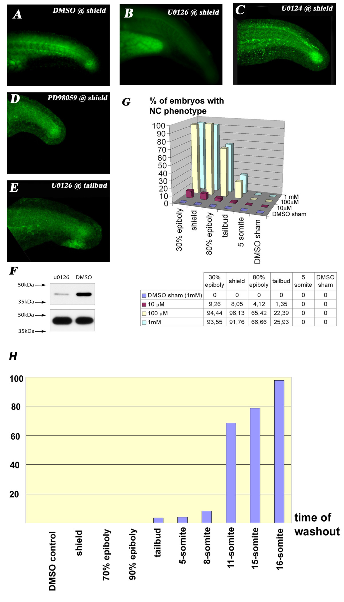
Fig. 1 U0126 application during a specific time-window decreases ERK phosphorylation specifically. A: Diphospho-Erk staining in the trunk of wt 24 hpf embryos.B: U0126 eliminates ERK1/2 phosphorylation (green) in the trunk and tail of 24 hpf zebrafish embryos. Notice that U0126-treated embryos have been overexposed (can be judged by the autofluorescence of the yolk sac extension) and still fail to show specific staining. C: U0124 does not eliminate the p-Erk staining. D: PD98059 also does not eliminate p-Erk staining when given just below the level of lethal toxicity (25 μM). E: Somewhat weaker but still significant p-Erk staining in embryos treated with U0126 starting at 10 hpf. F: Western-blot analysis demonstrated a slight but incomplete reduction in pErk1/2 levels in 10-somite embryos.G: Stage and dose-dependency of U0126 application. There is a dramatic drop in the percentage of affected embryos when treatment is applied after epiboly is completed. H: Washout experiments reveal the necessity of U0126 to be present until at least the 18 somite stage to produce the full phenotype.
Image
Figure Caption
Acknowledgments
This image is the copyrighted work of the attributed author or publisher, and
ZFIN has permission only to display this image to its users.
Additional permissions should be obtained from the applicable author or publisher of the image.
Full text @ BMC Dev. Biol.

