
|
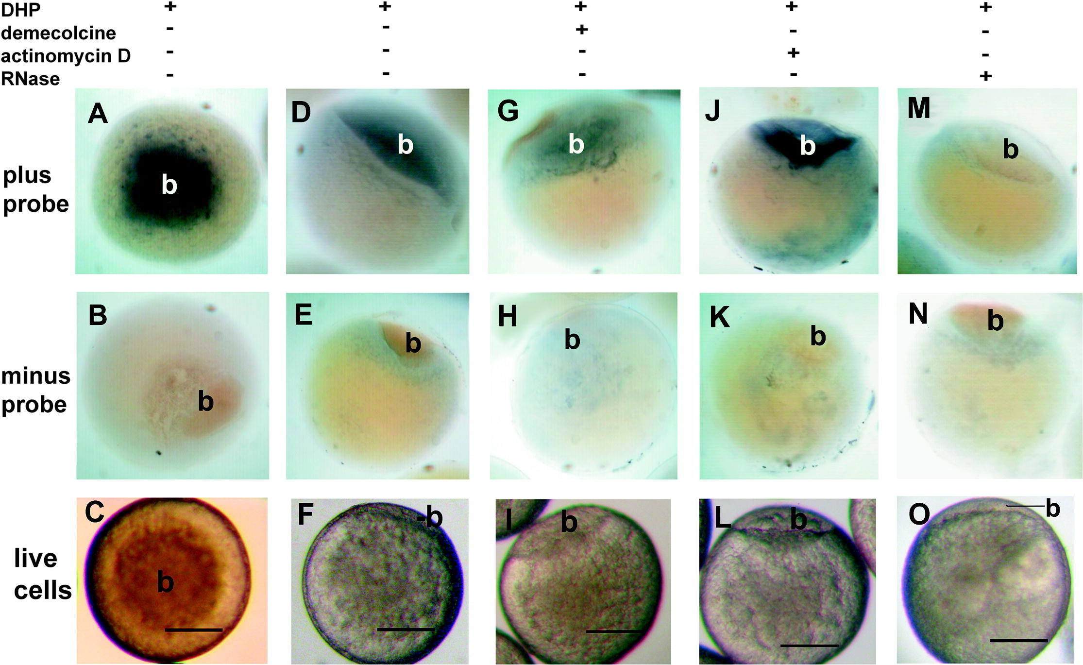
Fig. 2 γ-Tubulin in situ hybridization of 17 α 20 β dihydroxyprogesterone (DHP 1 μg/ml) matured oocytes. γ-Tubulin message was found predominately in the animal pole primordial blastodisc (b). (A, D, G, J and M) incubated with probe; (B, E, H, K, and N) incubated without probe. (M and N) preincubated with RNase prior to hybridization. (G and H) preincubated with demecolcine (1 μg/ml) and DHP prior to fixation and hybridization. J and K preincubated with actinomycin D (1 μg/ml) and DHP prior to fixation and hybridization. (C, F, I, L, and O) are live cells showing primordial blastodisc (b) formation at the animal pole after DHP incubation. (A, B and C) are animal pole views, while all others are side views. Scale bar equals 200 μm.
Reprinted from Gene expression patterns : GEP, 8(4), Liu, J., and Lessman, C.A., Changes of gamma-tubulin expression and distribution in the zebrafish (Danio rerio) ovary, oocyte and embryo, 237-247, Copyright (2008) with permission from Elsevier. Full text @ Gene Expr. Patterns