
|
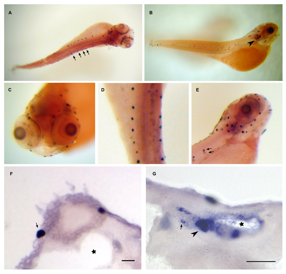
Fig. 13 Dr-S100T expression pattern by whole mount in situ hybridization. Five day old zebrafish larvae were hybridized with RNA antisense probe. Panels A) to E), whole mounts; panels F) to G), sectioned after hybridization. Scale bars 30 μm. Dorsal (A) and lateral (B) view show expression in lateral line neuromasts (arrows) and the otic placode (arrowhead). C) Frontal view of the head region. Several labeled neuromasts are visible. D) Magnification of the trunk region from panel A), view from dorsal. E) Enlarged lateral view of the head region, over ten labeled neuromasts are seen. One neuromast and its contra lateral counterpart are indicated by arrows.