
|
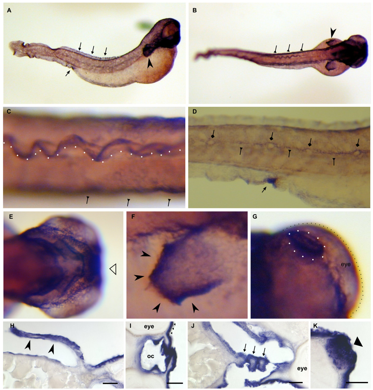
Fig. 11 Dr-S100I.1 expression pattern by whole mount in situ hybridization. Five day old zebrafish larvae were hybridized with RNA antisense probe. Panels A) to G), whole mounts; panels H) to K), sectioned after hybridization. Scale bars 30 μm. A) Lateral view, strong ubiquitous expression is seen in the skin, the urogenital opening (bottom arrow), the rim of the dorsal fin (top row of arrows), the pectoral fin (arrowhead) and the lower jaw. B) Dorsal view, strong expression in the pectoral fin, the dorsal fin, and the lateral line (triangle-headed arrows) C) Enlargement of dorsal view, expression in the lateral line (arrows) and the dorsal fin (rim indicated by white spots). D) Larger magnification of urogenital opening (arrow) and the labeled lateral line (triangle-headed arrows) surrounding the neuromasts (diamond-headed arrows), which are not labeled. E) Ventral view, strong expression in branchial arches is seen. F) Enlargement of pectoral fin, especially the rim is heavily labeled. G) Dorsal view, expression in the olfactory placode (white dots). H) Cross section of the pectoral fin. I) Pharynx is intensely labeled. J) Three branchial arches (asterisks) are cross-sectioned; the mesenchyme including the cartilaginous bar is devoid of staining. K) The olfactory placode is heavily stained.