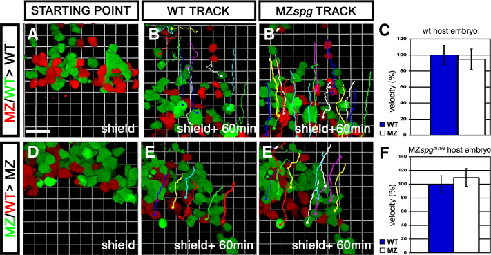
Fig. 6 Non-cell autonomous delay of DEL cell migration in MZspg embryos. Confocal z-stack time lapse recordings of mosaic embryos, starting at shield stage for 60 min. An approximately equal amount of cells from WT and MZspgm793 donors labeled with different fluorescent dyes were collected and co-transplanted into the ventral margin of a non-labeled host. Donors were either labeled with Alexa488-dextran (MW: 10 kDa) or Alexa546-dextran (MW: 10 kDa). (A, B) Cells transplanted into a WT host. (D, E) Cells transplanted into a MZspgm793 host. (B, E) Tracking lines of transplanted WT cells. (B′, E′) Tracking lines of transplanted MZspgm793 cells at the end of the time lapse. (C, F) Measurements of the net velocity of 10–20 transplanted cells. Velocity was normalized on WT velocity and depicted as % of WT data including standard deviation. (C) Net velocity of cells in a WT environment (n = 4): WT = 100 ± 12%; MZspgm793 = 95 ± 13%; p = 0.828. (F) Net velocity of cells in a pou5f1 deficient environment (n = 2): WT = 100 ± 22%; MZspgm793 = 110 ± 18%; p = 0.825. No significant difference in velocity was observed. Scale bar: 46.2 μm (Movies S1 and S2).
Reprinted from Developmental Biology, 315(1), Lachnit, M., Kur, E., and Driever, W., Alterations of the cytoskeleton in all three embryonic lineages contribute to the epiboly defect of Pou5f1/Oct4 deficient MZspg zebrafish embryos, 1-17, Copyright (2008) with permission from Elsevier. Full text @ Dev. Biol.

