
|
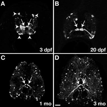
Fig. 1 Transverse sections of spinal cords of Tg(olig2:egfp) transgenic zebrafish, dorsal to top. Strong EGFP fluorescence was evident in ventral cells with radial processes extending to the pial surface at all stages (arrows). A: At 3 dpf, other EGFP+ cells had multiple fine membrane processes and occupied positions characteristic of OPCs (arrowheads). B-D: The number of EGFP+ radial cells in transverse sections remained constant at all stages examined through 3 months whereas the number of non-radial EGFP+ cells increased throughout the spinal cord. Scale bar = (A) 20 μM; (B) 30 μM; (C) 50 μM; (D) 80 μM.