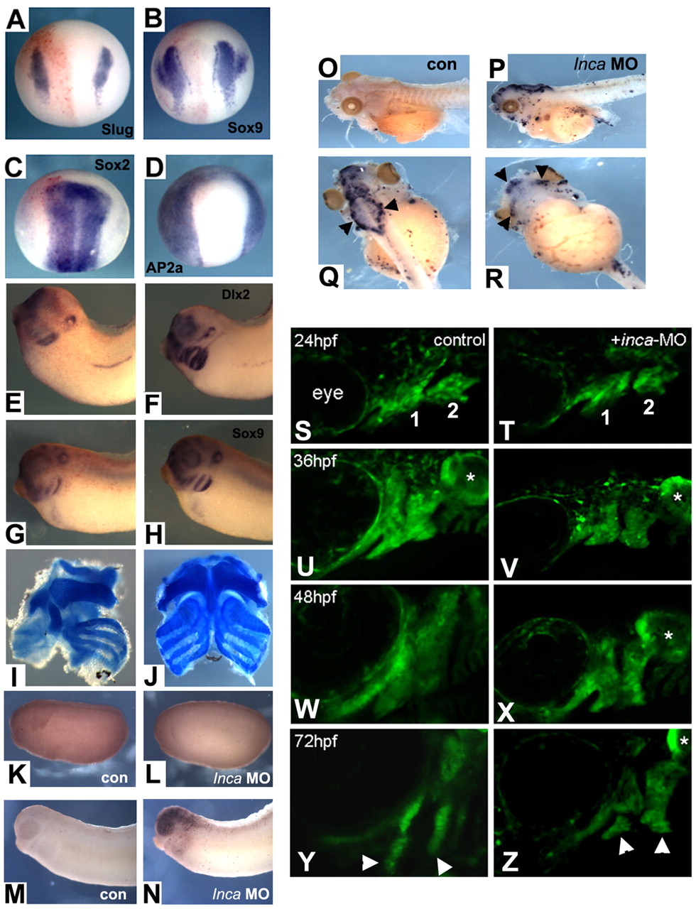
Fig. 5 Inca is required for cranial NC specification and survival after migration into the arches in Xenopus and zebrafish. Embryos were co-injected into one cell at the two-cell stage with Inca MO, together with lacZ RNA as a lineage tracer and assayed for gene expression by whole-mount in situ hybridization at stage 14 (A-D, injected sides are on the left) and stage 32 (E-H; E,G, injected sides) with probes indicated in the panels. In no case was any change in gene expression observed on the injected side [(A) Slug, n=83; (B) Sox9, n=42; (C) Sox2, n=47; (D) AP2a, n=54; (E) Dlx2, n=63; (G) Sox9, n=78]. At tadpole stage, cranial cartilage was eliminated on the injected side (left side in I). Control MO injection had no effect (J). (K-R) TUNEL staining of embryos injected with control MO (K,M,O) or Inca MO (L,N,P-R), cultured to stage 21 (K,L), stage 30 (M,N) or stage 45 (O-R). Q and R are dorsal and ventral views, respectively, of the tadpole shown in P. Black arrowheads indicate strong TUNEL signal in neurocranium (Q) and jaw cartilage (R). (S-Z) Confocal images of sox10:egfp expression in cranial NC cells of controls (S,U,W,Y) and inca1 morphants (T,V,X,Z). Lateral views, anterior to the left. Stages are indicated in the panels. First and second pharyngeal arches are indicated in S,T, and derived cartilage elements by arrowheads in Y,Z. Asterisks indicate expression of sox10:eGFP in the otic vesicle.
Image
Figure Caption
Figure Data
Acknowledgments
This image is the copyrighted work of the attributed author or publisher, and
ZFIN has permission only to display this image to its users.
Additional permissions should be obtained from the applicable author or publisher of the image.
Full text @ Development

