
|
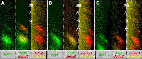
Fig. 5 Analysis of her1 and deltaC expression in staged embryos reveals deltaC expression in transition from anterior half-somite to posterior half-somite. (A–C) shows the right half of the trunk of three progressively older embryos. These embryos were fixed at the 10 somite stage, processed to visualize her1 mRNA (green), deltaC mRNA (red) and β-catenin protein (yellow). The embryos were more precisely staged by measuring the distances between the expression domains and the morphological somite borders. Embryos with decreasing distances between the expression domains and borders are considered progressively older (Holley et al., 2000). For each embryo, her1 expression is shown alone (left) or overlain with deltaC expression (middle). deltaC expression is also overlain with β-catenin localization (right). Note that since the somitic expression of deltaC is weak relative to the PSM expression, in the deltaC/β-catenin overlay, we have altered the levels of the only somitic expression in order to visualize this expression without saturating the PSM expression. The somite borders are highlighted (dashed lines).
Reprinted from Developmental Biology, 286(2), Julich, D., Hwee, Lim C., Round, J., Nicolaije, C., Schroeder, J., Davies, A., Geisler, R., Lewis, J., Jiang, Y.J., Holley, S.A., Tübingen 2000 Screen Consortium., beamter/deltaC and the role of Notch ligands in the zebrafish somite segmentation, hindbrain neurogenesis and hypochord differentiation, 391-404, Copyright (2005) with permission from Elsevier. Full text @ Dev. Biol.