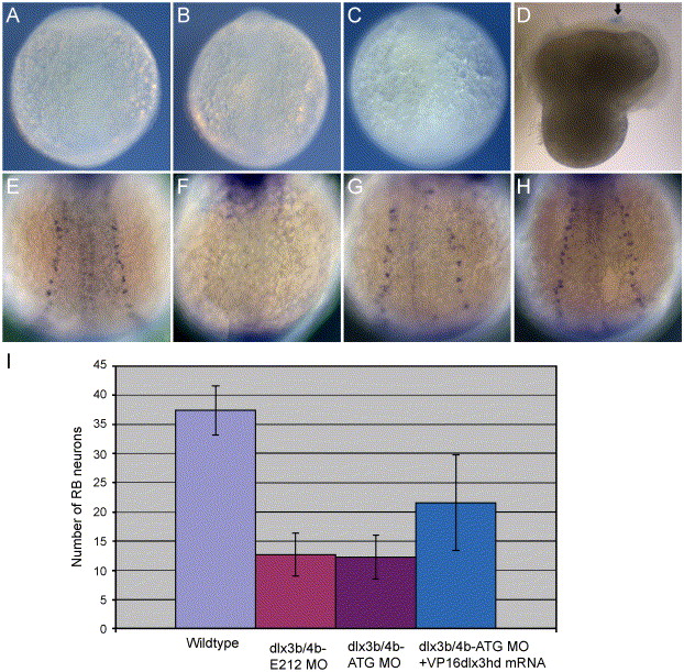
Fig. 6 Study of necrosis and rescued phenotypes of RB neurons by VP16-dlx3bhd mRNA in dlx3b and dlx4b MOs-injected embryos. Trypan blue-stained pattern in 2-3-somite-stage embryos (A–D) and HuC-expressing RB neuron pattern in 3-somite-stage embryos (E–H). (A) A wild-type embryo. (B) A 20-ng ATG-dlx3b-MO/20 ng ATG-dlx4b-MO-injected embryo. (C) A 40-ng control MO-injected embryo. (D) An embryo injured by a tungsten needle. No trypan blue-stained pattern is seen in wild type, 20 ng ATG-dlx3b-MO/20 ng ATG-dlx4b-MO-injected, and 40 ng control MO-injected embryos (A–C). In the injured embryo (D), a blue-stained pattern can be seen (arrow), indicating cell necrosis. (E) A wild-type embryo expressing HuC. (F) An embryo injected with 20 ng ATG-dlx3b-MO/20 ng ATG-dlx4b-MO. (G) A partially rescued embryo in RB neurons by VP16-dlx3bhd mRNA, injected with 20 ng ATG-dlx3b-MO/20 ng ATG-dlx4b-MO. (H) A fully rescued embryo in RB neurons by VP16-dlx3bhd mRNA, injected with 20 ng ATG-dlx3b-MO/20 ng ATG-dlx4b-MO. (I) Numbers of RB neurons are recovered in MO-injected embryos by injection of 150 pg VP16-dlx3bhd mRNA.
Reprinted from Developmental Biology, 276(2), Kaji, T., and Artinger, K.B., dlx3b and dlx4b function in the development of Rohon-Beard sensory neurons and trigeminal placode in the zebrafish neurula, 523-540, Copyright (2004) with permission from Elsevier. Full text @ Dev. Biol.

