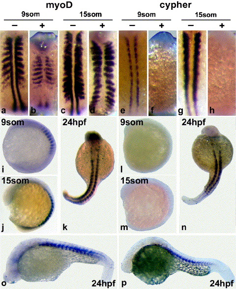
Fig. 7 Effect of cyclopamine treatment on cypher gene expression. Shown in a, c, e, and g are control treated embryos. All other embryos were incubated with 50 μM cyclopamine (starting at shield stage). The minus and plus refer to cyclopamine treatment but only for the first row embryos, which show the dorsal view of somites at the indicated stages. The left panel of embryos (a–d, i, j, k, and o) was probed for myoD expression. myoD expression was examined as a control and to show the absence of myoD expressing adaxial slow muscle precursor cells in the somite stage embryos upon cyclopamine treatment (b and d). The right panel embryos (e–h, l, m, n, and p) were probed for cypher expression. Cypher expression was absent in 9-somite and 15-somite stage embryos treated with cyclopamine (f and h). Embryos at 24 h of development showed slightly reduced cypher expression in paraxial somites when treated with cyclopamine. This is more apparent in the dorsal view (p). Normal expression of cypher and myoD during embryogenesis is shown in Fig. 2A. The control treatment with ethanol had no effect on either cypher or myoD expression. Similar results were obtained in three independent experiments.
Reprinted from Developmental Biology, 299(2), van der Meer, D.L., Marques, I.J., Leito, J.T., Besser, J., Bakkers, J., Schoonheere, E., and Bagowski, C.P., Zebrafish cypher is important for somite formation and heart development, 356-372, Copyright (2006) with permission from Elsevier. Full text @ Dev. Biol.

