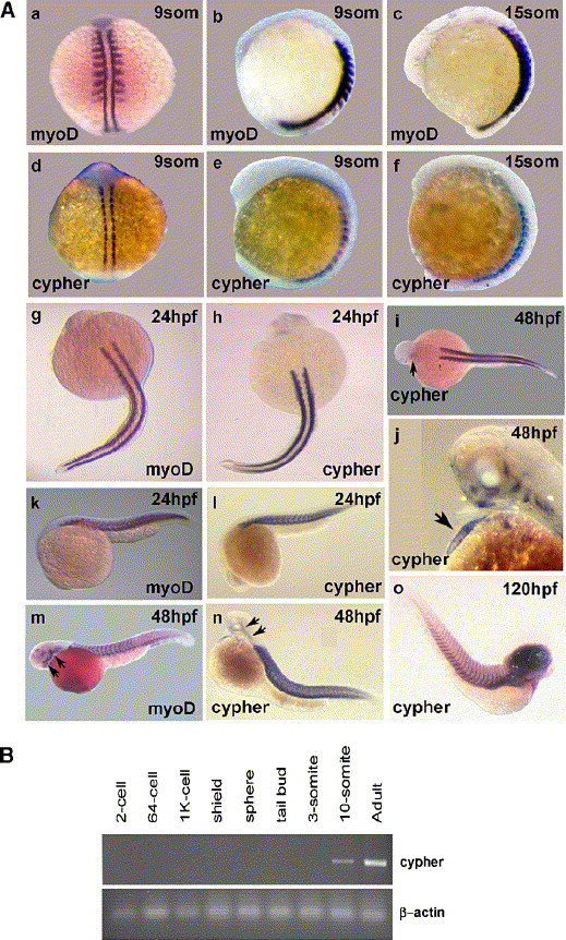
Fig. 2 Temporal and spatial expression of cypher in developing zebrafish. (A) Shown are whole mount in situ hybridization results for the skeletal muscle-specific marker myoD and for cypher at different stages of zebrafish development. The earlier 9-somite (9som) stage (14 hpf) and 15-somite stage (about 17 hpf) are shown in a–f. Shown are dorsal and lateral views. Limited cypher expression was already observed at the 3, 5, and 7 somite stages (see Supplementary Fig. 1). Embryos at 24 h of development (g, h, k, and l), 48 h of development (i, j, m, and n), and 120 h of development (o) are shown below. Used was either a myoD or a cypher specific probe, as indicated. The arrows in i and j indicate staining in the heart and in m and n point to head and jaw musculature. Staining in the heart was observed for cypher at 36 hpf (see Supplementary Fig. 1) but not at 24 hpf (h and l). Myod staining was not present in the heart at all times. Expression for both cypher and myoD was detected in head and jaw musculature at 48 hpf (m, n and also j). Cypher expression in the brain was detected at 120 hpf (o). (B) Semi-quantitative RT-PCR using cypher2γ designed primers and RNA from different stages of zebrafish development standard RT-PCR protocols were used. The PCR was done with 28 cycles and β-actin was used to control quality and quantity of RNA used for the different embryonic stages.
Reprinted from Developmental Biology, 299(2), van der Meer, D.L., Marques, I.J., Leito, J.T., Besser, J., Bakkers, J., Schoonheere, E., and Bagowski, C.P., Zebrafish cypher is important for somite formation and heart development, 356-372, Copyright (2006) with permission from Elsevier. Full text @ Dev. Biol.

