Fig. 5
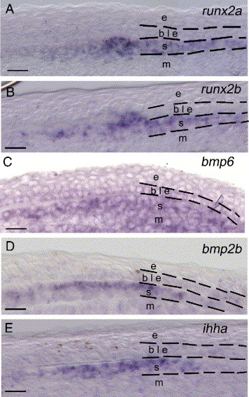
Fig. 5 Gene expression analysis using in situ hybridization on sections of 4 dpa regenerates. The pictures are focusing on a portion of the distal part of the regenerate taken at the same proximal–distal level and highlighting the common expression of runx2a, runx2b, bmp6, bmp2b and ihha in the newly differentiating scleroblasts. (A–B) The duplicate genes runx2a and runx2b are expressed in the differentiating scleroblasts in an overlapping expression pattern as seen on these adjacent sections (16 μm apart). (C) bmp6 is expressed in the differentiating scleroblasts, the mesenchymal cells of the blastema and a subset of cells of the basal epidermal layer. (D, E) bmp2b and ihha are similarly expressed within differentiating scleroblasts. Dashed lines separate different cell types within the fin regenerate. b, blastema; ble, basal layer of the epidermis; m, mesenchyme; s, scleroblasts. Scale bars: (A–E) 12.5 μm.
Reprinted from Developmental Biology, 299(2), Smith, A., Avaron, F., Guay, D., Padhi, B.K., and Akimenko, M.A., Inhibition of BMP signaling during zebrafish fin regeneration disrupts fin growth and scleroblast differentiation and function, 438-454, Copyright (2006) with permission from Elsevier. Full text @ Dev. Biol.

