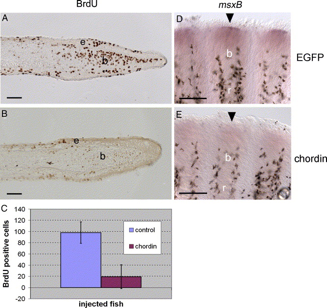
Fig. 3 Reduced cell proliferation and msxB expression following chordin construct injection. (A-B) Immunodetection of BrdU incorporation using an anti-BrdU antibody on longitudinal sections of fin rays at 4 dpa, 1 day after EGFP (A) and chordin (B) injections. Fish were treated with BrdU for 16 h starting immediately after injection. (C) For statistical purposes, BrdU labeled cells were counted on 20 sections of chordin-injected fin rays (n = 4 rays) and 9 sections of EGFP-injected rays (n = 2 rays) and the mean number of BrdU positive cells was calculated for both groups and plotted on a graph (Chordin = 19.4 cells, EGFP = 98.1 cells). Student′s t test results indicate that injection of chordin significantly decreased the number of proliferating cells within the blastema (P < 0.0001). (D-E) msxB gene expression on whole-mount fins at 4 dpa, 1 day after EGFP (C) and chordin (D) injections. msxB expression in the distal blastema (D, arrowhead) is strongly reduced in chordin-injected rays (E, arrowhead). b, blastema, e: epidermal layer, r: fin ray. Distal part of the regenerate is to the right (A, B) and to the top (D, E). Scale bars: (A, B) = 25 μm, (D, E) = 100 μm.
Reprinted from Developmental Biology, 299(2), Smith, A., Avaron, F., Guay, D., Padhi, B.K., and Akimenko, M.A., Inhibition of BMP signaling during zebrafish fin regeneration disrupts fin growth and scleroblast differentiation and function, 438-454, Copyright (2006) with permission from Elsevier. Full text @ Dev. Biol.

