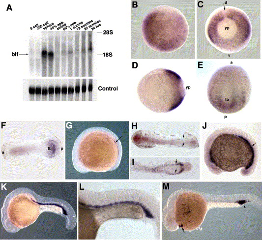
Fig. 2 Analysis of the blf expression pattern. (A) Northern blotting analysis of blf expression. A single blf-specific band of ∼2.4 kb size is observed from the sphere stage onwards. Maximum expression is observed at the sphere and 45% epiboly stages. 28S and 18S rRNA bands are indicated. (B–M) In situ hybridization analysis of blf expression. Anterior is to the left except where indicated. (B) 50% epiboly stage, animal view. blf expression appears uniform and ubiquitous within the blastoderm. (C, D) 80% epiboly stage, (C) vegetal view, (D) lateral view. blf is expressed within the posterior hypoblast and excluded from the dorsalmost cells (arrow, C). d, dorsal; v, ventral; yp, yolk plug. (E) 1-somite stage, dorso-posterior view. blf is localized to the posterior dorsal paraxial and lateral mesoderm and excluded from the axial mesoderm. Expression extends into the ventral region close to the tailbud (tb). a, anterior, p, posterior. (F) 3-somite stage, flat mount. blf is expressed in a semicircular pattern around the tailbud region (tb) in the dorsal paraxial and lateral mesoderm. a, anterior, p, posterior. (G, H) 10-somite stage. blf is expressed in two stripes of the putative hematopoietic progenitor cells in ventrolateral mesoderm (arrows). (I) 18-somite stage, the tail region. blf is localized to two stripes within the hematopoietic region of ventrolateral mesoderm. (J) 15-somite stage. blf is expressed in the presumptive hematopoietic cells in ventrolateral posterior mesoderm. (K, L) 22 hpf stage. blf is localized to the intermediate cell mass (ICM) region, where the primitive hematopoiesis is known to take place. (M) 26 hpf stage. blf is expressed in the ICM region (arrowhead) and circulating blood cells (arrows).
Reprinted from Developmental Biology, 283(1), Sumanas, S., Zhang, B., Dai, R., and Lin, S., 15-Zinc finger protein Bloody Fingers is required for zebrafish morphogenetic movements during neurulation, 85-96, Copyright (2005) with permission from Elsevier. Full text @ Dev. Biol.

