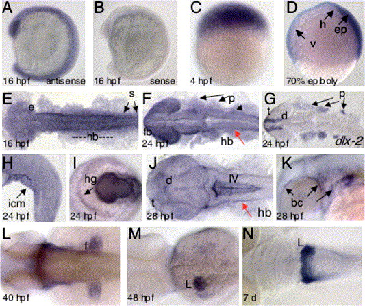
Fig. 2 sid4 expression during normal zebrafish development. (A) Embryos hybridized to sid4 antisense probe exhibited ubiquitous expression throughout early embryogenesis. (B) No signal was seen in embryos hybridized to the sense probe. (C) sid4 is expressed throughout the blastoderm at 4 hpf and (D) in the hypoblast (h), epiblast (ep), and ventral blastomeres (v) of gastrulating embryos. (E) sid4 is detected in the eye primordia (e), somites (s) and central nervous system of 16 hpf embryos. (F) sid4 is expressed in the forebrain (fb), hindbrain (hb), eyes, and pharyngeal arch region (p) of 24 hpf embryos. (G) dlx-2 expression is more restricted than sid4 within the pharyngeal arch. (H) The inner cell mass (icm) and (I) hatching gland (hg) of 24 hpf embryos also express sid4. (J) sid4 is absent from the 28-hpf hindbrain (red arrow) but prominently expressed in ventricle IV (IV), telencephalon (t), and diencephalon (d). (K) Scattered blood cells (bc) and (L) fin buds (fb) express sid4 at 40 hpf. (M) By 48 hpf, sid4 is restricted to the liver, where it remains through 7 days of development (N). Lateral views in A–D; H, K. Dorsal views in E–G; J–N. Anterodorsal view in I. Embryonic stages are indicated at the lower left of each panel.
Reprinted from Developmental Biology, 282(1), diIorio, P.J., Runko, A., Farrell, C.A., and Roy, N., Sid4: A secreted vertebrate immunoglobulin protein with roles in zebrafish embryogenesis, 55-69, Copyright (2005) with permission from Elsevier. Full text @ Dev. Biol.

