Fig. 3
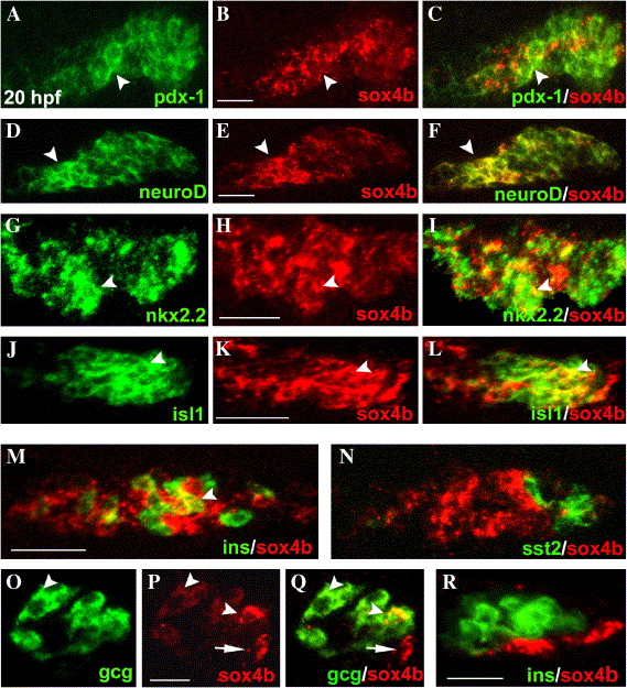
Fig. 3 Expression of sox4b in pancreas observed by FISH. (A–L) Expression of either pdx-1, neuroD, nkx2.2, isl1 (left column), or sox4b (central column) in 20 hpf embryos. The right column presents the overlay of the respective left and middle panels. (M, N) Overlay of sox4b- and either insulin- (M) or somatostatin2 (N) expression in 20 hpf embryos. (O, P) Expression of glucagon (O) and sox4b (P) in 36 hpf embryos. (Q) Overlay of panels O and P. (Q) Overlay of sox4b- and insulin expression in 26 hpf embryos. All the panels are optical sections of ventrally viewed yolk-free embryos (A–Q) and laterally viewed yolk-free embryo (R), with anterior to the left. sox4b was detected with a digoxigenin-labeled anti-sense probe and a tyramide-Cy3 substrate and the other markers with DNP-labeled anti-sense probes and tyramide-FITC substrate (see Materials and methods). A–N scale bars = 50 μm; O–Q scale bars = 25 μm. The arrowheads point to cells where sox4b is co-expressed with another marker, arrows in panels P and Q point to cells expressing only sox4b.
Reprinted from Developmental Biology, 285(1), Mavropoulos, A., Devos, N., Biemar, F., Zecchin, E., Argenton, F., Edlund, H., Motte, P., Martial, J.A., and Peers, B., sox4b is a key player of pancreatic alpha cell differentiation in zebrafish, 211-223, Copyright (2005) with permission from Elsevier. Full text @ Dev. Biol.

