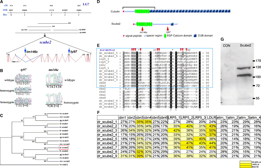
Fig. 3 Identification of Scube2 as the gene mutated at the you locus. (A) Positional cloning of the you locus. A critical region on linkage group 7 (LG7) was defined by simple sequence repeat marker (SSR) mapping and marker z11119 was found to be non-recombinant in 1400 meioses. A probe derived from the non-repetitive portion of z11119 detected PAC2D2. Further screening using PAC end cloning isolated BAC9A3. These two clones were shotgun sequenced and this sequence subsequently used to identify contiguous stretches of sequence in the emerging zebrafish genome sequence (ctg 08658, ctg 01991, ctg 09688,). The scube2 open reading frame was identified within this sequence, and the full genomic structure of the gene identified with reference to identified cDNA sequences. Intron sequences between exons 15 and 17 could not be defined due to gaps in the assembled genomic sequence. Arrow at exon 14 indicates the site of the identified stop codon in the youty97 allele. Arrow at exon 5 indicates the site of the identified stop codon in the youtm146c allele with the blue lines indicating the alternative splicing detected for this exon. (B) A stop codon is produced by mutation of a C to a T at the first base of codon Q644 and segregates with the youty97 phenotype. A stop codon in produced by mutation of a G to a T at the first base of codon E164 and segregates with the youtm146c phenotype. (C) Bootstrap phylogenetic analysis of all known Scube proteins, reveals that the you gene encodes a zebrafish protein orthologous to mammalian Scube2. The tree is rooted with human matrillin 2, the most closely related protein outside the Scube family. Abbreviations: hs, Homo sapiens; mm, Mus musculus; gg, Gallus gallus; dr, Danio rerio; tn, Tetraodon nigroviridis. (D) Structural homology of Cubilin and Scube2. The region sufficient for Cubilin binding to Megalin is bracketed. The position of the stop codon produced by the youty97 and youtm146c mutations is indicated by red arrows. (E) The EGF repeats of Scube2 and Cubilin share specific amino acid residues that define a separate Calcium binding EGF repeat class. Pan alignment of 73 seed sequences representative of all 5329 EGF domains present within PFAM and all EGF domains of the calcium binding class. This analysis reveals that specific EGF repeats (blue box) of zebrafish (dr) Scube2 (2, 3, 5, 6, 7, 9) share closest identity and specific residues (red arrow heads) with specific EGF repeats of human proteins, Cubilin (cbn), the LDL receptor (ldlr) and the LDLR related protein LRP-5. (F) Table of EGF domain homologies. A matrix of highest level homologies for the nine zebrafish Scube2 EGF domains reveals a separate sub-class of EGF containing proteins. Human Cubulin (cbn) and other endocytic receptors of the LDL receptor class (LRP5 and LDLR) share a high and distinct level of homology. The EGF repeats of human Attractin (attrn), which are not within this class, are included for comparison. (G) Western blot showing that zebrafish Scube2 is secreted from Scube2 transfected cells. Media from myc-tagged Scube2 transfected cells (Scube2) contains two secreted forms of zebrafish Scube2 protein, the larger at a similar molecular weight predicted by the full ORF and from similar experiments utilising the mammalian Scube proteins. These proteins are absent from media harvested from untransfected cells (CON). Marker sizes in kDa are shown.
Reprinted from Developmental Biology, 294(1), Hollway, G.E., Maule, J., Gautier, P., Evans, T.M., Keenan, D.G., Lohs, C., Fischer, D., Wicking, C., and Currie, P.D., Scube2 mediates Hedgehog signalling in the zebrafish embryo, 104-118, Copyright (2006) with permission from Elsevier. Full text @ Dev. Biol.

