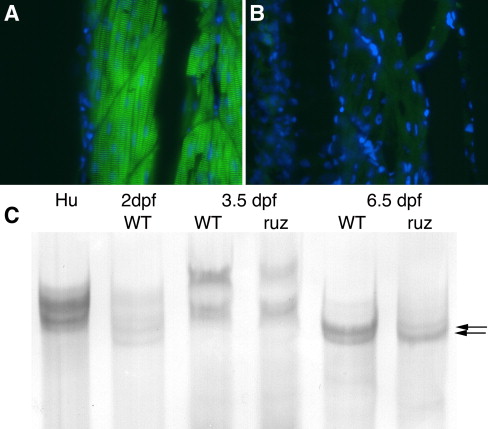
Fig. 4 ruz mutants exhibit decreased expression of specific titin isoforms. (A, B) Indirect immunofluorescence was performed on skeletal muscle longitudinal sections at 6 dpf. Nuclei were stained with DAPI (blue). (A) Wild-type muscle shows titin expression in a repeating sarcomeric pattern. (B) Mutant muscle shows severely decreased titin expression when imaged for the same exposure time. (C) Protein lysates of human psoas muscle (Hu), 2 dpf, 3.5 dpf, and 6.5 dpf wild-type (WT) zebrafish, and 3.5 dpf and 6.5 dpf ruz mutants (ruz) were separated by electrophoresis in adjacent wells of an SDS–agarose gel, transferred to nitrocellulose, and stained with India ink. Lysates loaded either by equal tissue weight per lysis volume or by equal total protein amount showed identical results. At 2 dpf, five faint titin isoforms are apparent. By 3.5 dpf, wild-type fish express four titin isoforms, including two isoforms with the lowest mobility seen during embryonic development. The low mobility doublet is slightly decreased in ruz mutants. At 6.5 dpf, wild-type zebrafish express two titin isoforms (arrows). The larger isoform is severely reduced in ruz mutants while the smaller variant appears increased. Identical results were seen in three independent experiments.
Reprinted from Developmental Biology, 309(2), Steffen, L.S., Guyon, J.R., Vogel, E.D., Howell, M.H., Zhou, Y., Weber, G.J., Zon, L.I., and Kunkel, L.M., The zebrafish runzel muscular dystrophy is linked to the titin gene, 180-192, Copyright (2007) with permission from Elsevier. Full text @ Dev. Biol.

