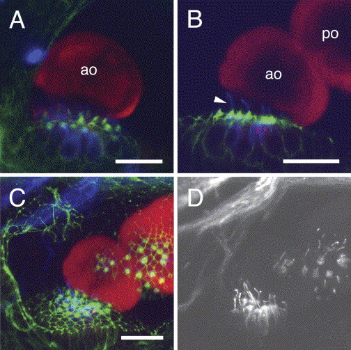
Fig. 6 Relationship of the fused otoliths in zotolin-1 MO-injected embryos to the corresponding sensory maculae. Triple-labeling with anti-OMP-1 (red), anti-acetylated tubulin (blue) antibodies, and phalloidin (green). Confocal fluorescence images at 72 hpf. Phalloidin (green) reveals the hair (stereocilia) bundles of the sensory cells and the contours of cells through their actin cortex. Acetylated tubulin (blue, false color) is found in the kinocilia and in the apical cytoplasm of the sensory hair cells (Riley et al., 1997). (A) Anterior macula and otolith of a control MO-injected embryo, lateral view. (B–D) zotolin-1 MO-injected embryo, with fused otoliths. (B) and (C) are the same z-stacks (3D reconstruction available upon request). (B) Anterior macula and anterior otolith with the fused posterior otolith; note the long kinocilia (arrowhead). (C) Projection of the confocal series along the z-axis (22 images, taken every 2 μm). Both maculae are visible through their hair bundles, in relation to both otoliths. (D) Far-red (blue-coded in A–C) fluorescence channel extracted from the image shown in (C), to show the acetylated tubulin immunostaining of kinocilia at both maculae. ao, anterior otolith; po, posterior otolith. Dorso-lateral view, anterior to the left, dorsal to the top. Scale bars, 20 μm.
Reprinted from Mechanisms of Development, 122(6), Murayama, E., Herbomel, P., Kawakami, A., Takeda, H., and Nagasawa H., Otolith matrix proteins OMP-1 and Otolin-1 are necessary for normal otolith growth and their correct anchoring onto the sensory maculae, 791-803, Copyright (2005) with permission from Elsevier. Full text @ Mech. Dev.

