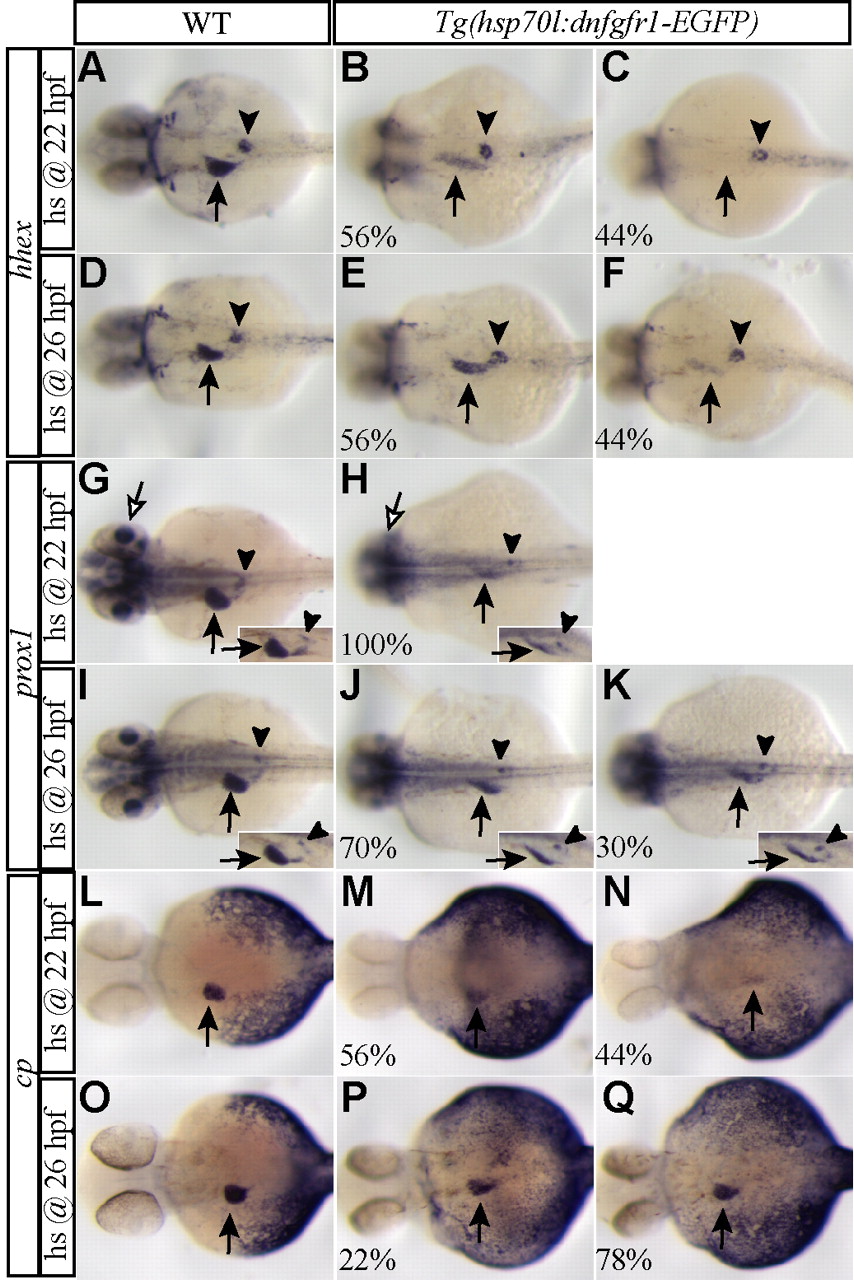
Fig. 7 Fgf signaling is not essential for the maintenance of specified liver progenitors. Embryos obtained from outcrossing a hemizygous Tg(hsp70l:dnfgfr1-EGFP) zebrafish were heat shocked at 22 (A-C,G,H,L-N) or 26 (D-F,I-K,O-Q) hpf, harvested at 35-36 (A-K) or 38-40 (L-Q) hpf and examined for hhex (A-F), prox1 (G-K), and cp (L-Q) expression. The percentage of the hemizygous embryos exhibiting a similar expression pattern is indicated in the lower left corner (n=8-10). When embryos were heat shocked at 22 hpf, hhex expression in the liver region (arrows) was greatly reduced (B) or almost absent (C) in the hemizygous embryos, whereas its expression in the pancreatic islet (arrowheads) appeared unaffected (B,C). prox1 expression in the liver (black arrows) and retina (white arrows) was also greatly reduced in the hemizygous embryos (H), whereas its expression in the interrenal primordium appeared unaffected (H, arrowheads). By contrast, when embryos were heat shocked at 26 hpf, both hhex and prox1 were clearly expressed in the liver region in all the hemizygous embryos (E,F,J,K, arrows). However, hhex and prox1 expression in the liver region was reduced compared with wild-type siblings, whereas hhex expression in the pancreatic islet and prox1 expression in the interrenal primordium appeared unaffected (E,F,J,K, arrowheads). Hepatocyte differentiation, assessed by cp expression, was severely defective in the hemizygous embryos heat shocked at 22 hpf (M,N, arrows) and weakly reduced in those heat shocked at 26 hpf (P,Q, arrows). All images, except insets, are dorsal views, anterior left. Insets are side views, anterior left.
Image
Figure Caption
Figure Data
Acknowledgments
This image is the copyrighted work of the attributed author or publisher, and
ZFIN has permission only to display this image to its users.
Additional permissions should be obtained from the applicable author or publisher of the image.
Full text @ Development

