
|
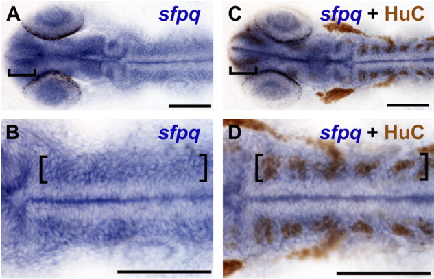
Fig. 4 sfpq is strongly expressed in regions of neurogenic activity. A-D: At 24 hours postfertilization (hpf) wild-type embryos labeled for sfpq expression by in situ hybridization only (A,B) and wild-type sibling embryos double labeled for sfpq mRNA expression and HuC protein by immunohistochemistry (C,D) show that regions of strongest sfpq expression in the brain (brackets) overlap with HuC, a marker for postmitotic neuronal precursors. B,D: Higher magnification of hindbrain; brackets mark strong sfpq expression overlapping with HuC labeling. Midline staining is an artifact of the staining process, because it is not observed in embryos cut open before staining. A-D: Dorsal views. Anterior left. Scale bar = 100 μm.