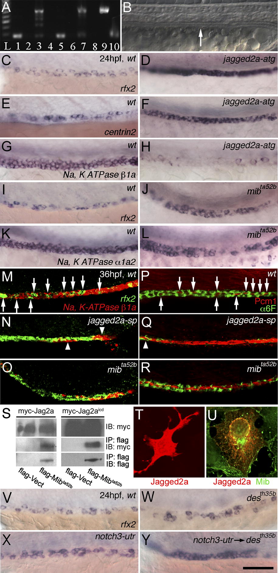
Fig. 4 Multi-Cilia Cell Hyperplasia Is Due to Mib-Mediated Jagged2a Signaling Pathway via Notch1a and Notch3 Receptors(A) Effectiveness of splicing jagged2a-sp MO. RT-PCR of control embryos generates a 230-bp jagged2a fragment, bridging parts of exon 1 and exon 2 at 24 hpf (lane 1) and 48 hpf (lane 5). jagged2a-sp MO-injected embryos analyzed with the same primers at the same timepoints (lanes 3 and 7) show a larger amplicon of 708 bp caused by a nonsplicing intron 1, which encodes a premature stop codon. Lane 9 shows the amplicon from genomic DNA, and lane 10 shows the amplicon from jagged2a cDNA. No fragment can be amplified in the RT-PCR without reverse transcriptase in 24-hpf (lane 2) or 48-hpf (lane 6) wt embryos or in 24-hpf (lane 4) or 48-hpf (lane 8) jagged2a-sp MO-injected embryos. Lane L: 100-bp ladder. (B) Pronephric duct (arrow) integrity is not affected in jagged2a morphants. Panels C–L focus on the duct between somite 10 and 13. (C–H) Multi-cilia cell number is increased in (D and F) jagged2a-atg morphants compared to (C and E) wt embryos as shown by (C and D) rfx2 and (E and F) centrin2 expression at 24 hpf, but principal cell number is decreased in (H) jagged2a-atg morphants compared to (G) wt embryos as revealed by Na+, K+ ATPase β1a expression at 24 hpf. (I–L) Multi-cilia cell number is increased in (J) mibta52b embryos compared to (I) wt embryos as shown by rfx2 expression at 24 hpf, but principal cell number is decreased in (L) mibta52b embryos compared to (K) wt embryos as revealed by Na+, K+ ATPase α1a2 expression at 24 hpf. Panels M–R focus on the duct around somite 11 to 13. (M–O) Fluorescent double in situ hybridization of rfx2 (green) and Na+, K+ ATPase β1a (red) in 36-hpf (M) wt embryos, (N) jagged2a-sp morphants, and (O) mibta52b mutants shows multi-cilia cell hyperplasia in jagged2a morphants and mibta52b mutants. Arrows point to the rfx2-expressing cells in the duct of (M) wt embryos; arrowheads point to the Na+, K+ ATPase β1a-expressing cells in the pronephric duct of (N) jagged2a-sp morphants. (P–R) Double immunohistochemistry of α6F (green) and Pcm1 (red) in 36-hpf (P) wt embryos, (Q) jagged2a-sp morphants, and (R) mibta52b mutants shows multi-cilia cell hyperplasia in jagged2a morphants and mibta52b mutants. Arrows point to the Pcm1 staining in the pronephric duct of (P) wt embryos; arrowheads point to α6F staining in the pronephric duct of (Q) jagged2a-sp morphants. (S) Immunoprecipitation of Myc-Jagged2a and Myc-Jagged2aicd by Flag-Mibta52b. IP, immunoprecipitation; IB, immunoblotting. (T–U) Expression of Myc-Jagged2a (T) and cotransfection of Myc-Jagged2a and Flag-Mib (U) in COS7 cells. (V–Y) Compared to (V) wt embryos, mild cilia cell hyperplasia is observed in (W) notch1a (desth35b) mutants and (X) notch3-utr morphants, while severe cilia cell hyperplasia is observed in (Y) notch3-utr MO-injected notch1a (desth35b) mutants as shown by rfx2 expression at 24 hpf. All embryos, anterior to the left. Bar scale: 100 μm (B), 75 μm (C–L and V–Y), 50 μm (M–R), and 30 μm (T and U).
Image
Figure Caption
Figure Data
Acknowledgments
This image is the copyrighted work of the attributed author or publisher, and
ZFIN has permission only to display this image to its users.
Additional permissions should be obtained from the applicable author or publisher of the image.
Full text @ PLoS Genet.

