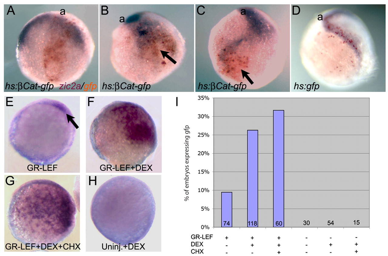
Fig. 5 Ectopic activation of the canonical Wnt pathway induces zic gene transcription. (A-C) Embryos were injected with hs:ßCat-gfp DNA, heat shocked at shield-stage, and assayed for zic2a (purple) and gfp (orange) at late-gastrula. (A) ßCat-gfp is expressed in the medial neural plate; the zic2a pattern is normal (no ectopic expression; n=10, two experiments). (B,C) ßCat-gfp is expressed in lateral ectoderm; zic2a is ectopically expressed (arrows; n=8, 8 ectopic, two experiments). (D) Control embryo expressing hs:gfp in lateral ectoderm showing no ectopic zic2a expression (n=18, 0 ectopic, one experiment). a, anterior. (E-G) Zic2aD5:gfp embryos injected with GR-Lef RNA and assayed for gfp RNA at late-gastrula (purple). (E) GR-Lef-injected embryo showing weak ectopic gfp (arrow). (F,G) GR-Lef-injected embryos treated with DEX (F) or DEX and CHX (G). (H) Uninjected zic2aD5:gfp control treated with DEX. (I) Summary of GR-Lef overexpression experiments, showing the proportion of embryos expressing gfp for each treatment condition. The number of embryos scored for each condition is listed along the x-axis.
Image
Figure Caption
Acknowledgments
This image is the copyrighted work of the attributed author or publisher, and
ZFIN has permission only to display this image to its users.
Additional permissions should be obtained from the applicable author or publisher of the image.
Full text @ Development

