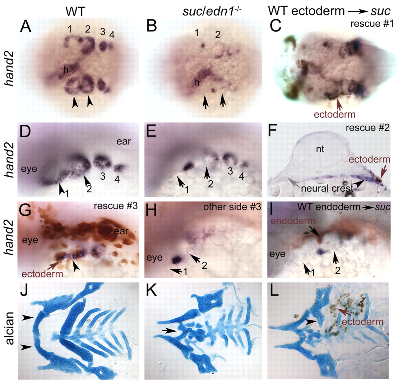
Fig. 7 Facial ectoderm is a crucial functional source of Edn1 in the arches. Whole-mount RNA in situ hybridizations at 30 hpf (A,B,D,E), with immunohistochemistry for biotin-dextran (brown cells in C,F-I). Dorsal views (A-C), dorsolateral views (D,E,G-I), 96 hpf flat-mounted Alcian-Blue-stained cartilages (J,K) combined with immunohistochemistry for biotin-dextran (L). (A,D) hand2 expression in ventral cranial NC cells in wild type (arrowheads), which is lost in suc;edn1-/- (B,E, arrows), except in few cells at arch borders. (C) Rescue of hand2 in suc;edn1-/- by unilateral grafting of ectoderm on the left side (arrow). (F) Transverse cryosection through the arch shows unilateral rescue of hand2 (arrowhead) in a suc;edn1-/- embryo adjacent to grafted ectoderm (brown cells, arrow). The control side did not receive any donor ectoderm and shows no rescue of hand2 (gray arrow). (G) Another example of wild-type ectoderm (brown cells, arrow) rescuing hand2 (arrowheads). (H) Control side of embryo in G. (I) Wild-type endoderm (brown cells, arrow) did not rescue hand2. (J) Wild-type cartilages include the mandibular, hyoid and branchial elements including Meckel's cartilage (arrowhead). (K) Meckel's cartilage is reduced in suc;edn1-/- (arrow). (L) suc;edn1-/- mutant that received wild-type ectoderm shows unilateral rescue of the ventral hyosymplectic cartilage (arrowhead) adjacent to the grafted ectoderm (arrow, brown cells). h, heart; nt, neural tube.
Image
Figure Caption
Figure Data
Acknowledgments
This image is the copyrighted work of the attributed author or publisher, and
ZFIN has permission only to display this image to its users.
Additional permissions should be obtained from the applicable author or publisher of the image.
Full text @ Development

