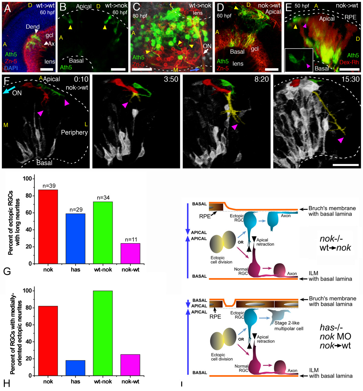
Fig. 10 Importance of the tissue environment for the differentiation and orientation of ectopic retinal ganglion cells (RGCs). (a) Extended-focus confocal image from a wild-type host embryo transplanted with blastomeres from an ath5:gap-gfp transgenic wild-type embryo. gcl, ganglion cell layer. (b,c) Extended-focus images from nok-/- host retinas transplanted with ath5:gap-gfp transgenic cells (wild type). In (b), the lateral view of the eye shows the presence of ectopic ath5:gap-gfp-positive cells (arrowheads); in (c), the eye is seen from a ventral view, and many ectopic (apical) donor RGCs that extend axons (yellow arrowheads) joining the optic nerve (ON) on the outside of the eye are shown. (d-f) Images of wild-type hosts transplanted with blastomeres from nok-/-, ath5:gap-gfp transgenic donors. (d) Extended-focus confocal image, in which the donor RGCs (positive for both Zn-5 (red) and ath5:gap-gfp (green)) are found either mixed with host RGCs in a normal-appearing ganglion cell layer (gcl) or on the apical side of the retina. (e) Rotated extended-focus confocal image, showing how a nok-/- donor-derived ectopic (apical; yellow arrowhead) ath5:gap-gfp-positive cell starts to extend a neurite (purple arrowhead) that will grow only a few micrometers before turning back to the originating cell (Additional file 10). The red stain shows all the donor cells, labeled with dextran-tetramethylrhodamine β-isothiocyanate (Dex-Rh). No transplanted cells are detected in the surrounding retinal pigment epithelium, indicating that it must all be of wild-type (host) origin. The inset shows a better view (from the apical side, in this case in an optical section orthogonal to the confocal laser) of the cell in the same time point. (f) Sequence of rotated three-dimensional reconstructions taken from a four-dimensional analysis. The two cells highlighted are from the donor (nok-/-) and are extending long neurites, which will grow together towards the retinal periphery (colored yellow where they are indistinguishable from each other). Time point 0 is at 32 hpf. ON, optic nerve; A, anterior; D, dorsal; L, lateral; M, medial. All scale bars are 25 μm long. (g) Comparison of the number of apically located ath5:gap-gfp cells extending a long neurite (longer than three cell diameters), in different mutant and morphant conditions. Total number of cells (n) and embryos analyzed (cells/embryos): nok-/-, 39/11; has-/-, 29/8; transgenic nok-wild-type, 34/9; transplanted wild-type-nok, 11/6. (h) The proportion of these cells in which the neurite is growing towards the medial region of the eye, where they would be able to meet the optic nerve. (i) Summary of the observed phenotypes of ectopic RGCs in mutant, morphant and transplantation conditions.
Image
Figure Caption
Acknowledgments
This image is the copyrighted work of the attributed author or publisher, and
ZFIN has permission only to display this image to its users.
Additional permissions should be obtained from the applicable author or publisher of the image.
Full text @ Neural Dev.

