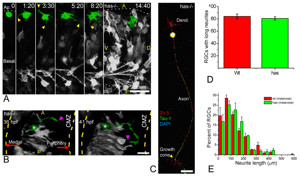
Fig. 8 Polarization of ectopic RGCs is impaired in a non cell-autonomous manner in has mutants. (a) Four-dimensional (4D) analysis of the retina of a has-/- embryo injected with ath5:gap-gfp plasmid DNA (Additional file 9). The cell highlighted in green divides at the first time frame, and the two daughter cells remain for several hours at the apical region of the retina, increasing their GFP expression but not generating any long neurites. Time point 0 is at 32 hpf. A, anterior, D, dorsal; P, posterior; V, ventral. Scale bar, 25 μm. (b) A dorsal view of a three-dimensional reconstruction taken from a 4D confocal analysis of a has-/- embryo transgenic for ath5:gap-gfp. The ectopic retinal ganglion cell (RGC) highlighted in green is growing a long neurite (arrowheads) that is initially directed towards the retinal periphery (that is, opposite to the optic nerve exit), and then turns back towards the cell. CMZ, ciliary marginal zone. Scale bar, 15 μm. (c) has-/- RGC in culture, triple-labeled with Zn-5 and anti-Tau-1 antibodies and 4',6-diamidino-2-phenylindole. The cell is indistinguishable from a stage 4 wild-type RGC (see Figure 1). Scale bar, 15 μm. (d,e) Quantitative analysis of neuritic outgrowth in vitro from has-/-, compared with wild-type, RGCs (defined as Zn-5-positive cells). (d) Number of RGCs with long (more than three cell diameters) neurites; n = 200 cells per strain from three independent experiments. (e) Distribution of neurite lengths; n = 100 neurites per strain (one neurite per cell; the longest was chosen in cells with more than one) from three independent experiments (normalized values).
Image
Figure Caption
Acknowledgments
This image is the copyrighted work of the attributed author or publisher, and
ZFIN has permission only to display this image to its users.
Additional permissions should be obtained from the applicable author or publisher of the image.
Full text @ Neural Dev.

