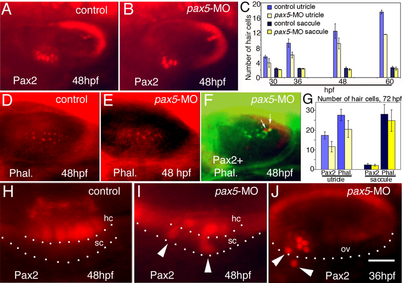
Fig. 5 Assessment of hair cell development. A,B: Anti-Pax2 staining in the otic vesicle in a control embryo (A) and pax5 morphant (B) at 48 hours postfertilization (hpf). C: Number of Pax2+ hair cells at the indicated stages (means and standard errors of at least three experiments, with at least 15 specimens/time-point/experiment). P values for the comparison of control embryos vs. pax5 morphants are as follows: utricle, P = 0.042 (30 hpf), P = 0.009 (36 hpf), P = 0.0007 (48 hpf), P = 0.017 (60 hpf); and saccule, P = 0.136 (30 hpf), P = 0.138 (36 hpf), P = 0.05 (48 hpf), P = 0.28 (60 hpf). D,E: Rhodamine-phalloidin staining in the saccular macula of a control embryo (D) and pax5 morphant (E) at 48 hpf. In control embryos (n = 23), there were 21.5 ± 5.0 hair cells in the utricular macula and 17.8 ± 9.7 in the saccular macula. In pax5 morphants (n = 42), there were 16.8 ± 4.5 hair cells in the utricle and 14.8 ± 7.5 in the saccule. F: Saccular maculae stained with anti-acetylated tubulin (green) and anti-Pax2 (red) in a pax5 morphant at 48 hpf. Only two hair cells are Pax2-positive (arrows). G: Hair cell numbers detected by anti-Pax2 or phalloidin staining in pax5 morphants and uninjected controls at 72 hpf. Data bars are color-coded as in C. H,I: Enlarged view of the utricular macula stained with anti-Pax2 at 48 hpf in a control embryo (H) and pax5 morphant (I). Basal edges of hair cell (hc) and supporting cell (sc) layers are indicated. J: Otic vesicle of pax5 morphant stained with anti-Pax2 at 36 hpf. Arrowheads mark misplaced hair cells. The ventral limit of the otic vesicle (ov) is indicated. Images show dorsolateral (A,B,D-F,J) and lateral (H,I) views with anterior to the left. Scale bar = 40 μm in A,B,D-F, 12.5 μm in H,I, 25 μm in J.
Image
Figure Caption
Figure Data
Acknowledgments
This image is the copyrighted work of the attributed author or publisher, and
ZFIN has permission only to display this image to its users.
Additional permissions should be obtained from the applicable author or publisher of the image.
Full text @ Dev. Dyn.

